MKA-chirurgie
Uitgangsvraag
Voor welke toepassingen binnen de mondziekten, kaak- en aangezichtschirurgie kunnen 3D-prints worden ingezet?
Aanbeveling
Aanbeveling-1
Denk aan het gebruik van 3D-prints binnen de mondziekten, kaak- en aangezichtschirurgie, specifiek voor de volgende hulpmiddelen:
- Anatomische modellen.
- Buigmodellen of -mallen.
- Chirurgische mallen.
- Splints.
- Patiëntspecifieke implantaten.
- Scaffolds.
Aanbeveling-2
Denk aan het gebruik van 3D-prints binnen de mondziekten, kaak- en aangezichtschirurgie in de volgende deelgebieden:
- Implantologie.
- Orthognathe Chirurgie.
- Oncologie.
- Aangezichtstraumatologie.
- Botaugmentatie.
Denk aan 3D-prints bij reconstructie van het cranium of neus, genioplastiek of TMJ-gewrichtsprothesevervanging.
Overwegingen
Balans tussen gewenste en ongewenste effecten
Conclusies uit de literatuur
De werkgroep heeft onderzocht welke 3D-prints binnen de Mondziekten, kaak- en aangezichtschirurgie (MKA) worden genoemd in wetenschappelijke publicaties. In deze review zijn de abstracts van 158 studies geanalyseerd. De literatuur over 3D-printen binnen de MKA laat zien dat deze techniek wijdverspreid wordt ingezet in het vakgebied: op het gebied van implantologie, orthognathe chirurgie, aangezichtstraumatologie, oncologie en botaugmentatie zijn meerdere gerandomiseerde controlestudies en systematic reviews naar 3D-geprinte hulpmiddelen binnen de patiëntenzorg gepubliceerd.
Uit de abstracts is niet altijd op te maken welk type 3D-print wordt geëvalueerd en welke type prints worden gecombineerd (bijvoorbeeld surgical guides met patiëntspecifieke implantaten). Aangezien niet benoemde 3D-prints ook niet zijn meegenomen in deze analyse, is het aannemelijk dat de werkelijke getallen rondom het gebruik van 3D-geprinte hulpmiddelen hoger zal liggen. Verder valt op dat in gerandomiseerde controlestudies waarin het gebruik van patiëntspecifieke implantaten wordt geëvalueerd een controlegroep wordt gebruikt die ook gebruik maakt van een op 3D-prints gebaseerde aanpak. Dit laat zien dat de inzet van 3D-geprinte hulpmiddelen inmiddels als standaardzorg wordt gezien.
De beschreven uitkomsten in de literatuur richten zich voornamelijk op chirurgische nauwkeurigheid. Hierover kan de conclusie worden getrokken dat de chirurgische nauwkeurigheid toeneemt wanneer 3D-printing tijdens de behandeling wordt ingezet. Functionele uitkomstmaten, patiënt gerapporteerde uitkomstmaten of operatieduur zijn op specifieke subgebieden – en soms zelfs alleen voor specifieke ingrepen binnen deze subgebieden – geanalyseerd. Hoewel ook deze resultaten positief zijn, gelden deze bevindingen slechts voor het subspecialisme of de ingreep die beschreven is en kan dit niet zomaar geëxtrapoleerd worden naar het gehele vakgebied vanwege de verschillen in indicatie en type ingreep. Andere uitkomstmaten zoals kosten zijn weinig beschreven en lijken te verschillen per type hulpmiddel, waardoor ook hierover geen conclusies kunnen worden getrokken aan de hand van de onderzochte literatuur.
Conclusies uit aanvullende onderzoek
Uit de enquête naar 3D-printen in Nederland, zie “Bijlage Uitwerking van de enquêteresultaten”, volgt dat de MKA niet alleen het langst gebruik maakt van 3D-printen (sinds 2006), maar ook de grootste aantallen 3D-prints gebruikt per jaar ten opzichte van andere medisch specialismen. Dit geldt voor alle typen 3D-prints; anatomische modellen, schedelmodellen, boor-/zaagmallen, implantaten, wafers/splints, patiëntspecifieke implantaten of reconstructiematerialen en wekedelenprints. De MKA geeft specifiek aan dat 3D-prints veel gebruikt worden bij het plannen van een operatie en dat kwaliteitsverbetering van de zorg en voorspelbaarder werken redenen zijn om met 3D-prints te werken.
In de MKA worden anatomische modellen gebruikt als zichtmodellen van complexe anatomie en pathologie, buigmodellen voor het aanbuigen van osteosynthesemateriaal en als pasmallen bij autotransplantaties. Verder wordt 3D-printing ook ingezet voor het vervaardigen van fysieke tandmodellen, als alternatief voor gipsmodellen.
De MKA is de grootste gebruiker van boor- en zaagmallen. Als toepassing van boor- en zaagmallen worden genoemd: uitvoeren van osteotomieën, oncologische resecties en reconstructies, plaatsing implantaten, orthognathe chirurgie, genioplastiek, en bij volledige vervanging van een kaakgewricht.
Volgens de ingevulde enquête voor de medisch specialisten maken de specialismen MKA (net als tandheelkunde) veelvuldig gebruik van splints en wafers. Deze worden als positioneringsmal gebruikt bij orthognathe chirurgie, waarbij de geplande verplaatsing van de dentitie van boven- en/of onderkaak en de geplande occlusie kunnen worden overgebracht naar de chirurgische setting. Verder worden splints ingezet als beschermplaatje na (oncologische) palatumchirurgie of maxillectomie, bij temporomandibulaire dysfunctie, als beschermplaat bij bruxisme. Door de respondenten van de enquête wordt aangegeven dat alle wafers door het eigen 3D-lab ontworpen worden. Op één na printen alle zes 3D-labs op hun eigen printer wafers voor de MKA.
De enquête geeft verder inzicht in het gebruik van implantaten binnen de MKA: de MKA gebruikt patiëntspecifieke implantaten of reconstructiematerialen zoals cranioplastiek implantaten en onbelaste craniofaciale implantaten. Daarnaast worden 3D-prints in de MKA gebruikt bij wekedelen-reconstructies.
Tevens is een enquête met vragen over het gebruik van 3D-prints in de klinische praktijk verstuurd naar patiënten. De methode en resultaten uit de patiënten enquête zijn te lezen in “Bijlage Patiëntenperspectief over gebruik 3D-model prints in de klinische praktijk" De resultaten uit de patiënten enquête schetsen een patiëntenperspectief waarin begrijpelijkheid, transparantie en contextgerichte communicatie centraal staan. Patiënten staan overwegend open en positief tegenover het gebruik van 3D-prints in de klinische praktijk, maar ervaren deze technologie vaak als weinig zichtbaar of onvoldoende toegelicht. Wanneer 3D-prints wél expliciet worden ingezet en besproken, waarderen patiënten deze vooral als hulpmiddel om hun aandoening, behandeling en keuzes beter te begrijpen. De meerwaarde wordt daarmee primair gezien op het niveau van uitleg, visualisatie en ondersteuning van besluitvorming, en minder als een direct middel om klinische uitkomsten te verbeteren. Dit samenvattende beeld onderstreept dat voor patiënten niet de technologie zelf, maar hoe deze wordt gebruikt en gecommuniceerd, bepalend is voor de ervaren waarde.
Algemene overwegingen
Over het algemeen worden positieve bevindingen gerapporteerd in de literatuur wanneer 3D-printing in de behandeling van de MKA-patiënt wordt ingezet. De respondenten van de enquête geven als motivatie om met 3D-prints te werken: kwaliteitsverbetering van zorg, verbeterde operatieplanning en voorspelbaarder werken. Het gebruik zoals beschreven in de enquêteresultaten lijkt overeen te komen met het overzicht vanuit de literatuur gegenereerd. Het printen van tandmodellen is de een toepassing die in de enquête naar voren komt, maar niet uit de literatuursearch. Met deze toevoeging geeft de informatie uit literatuur en enquête een goed totaalbeeld van het gebruik binnen de mondziekten, kaak- en aangezichtschirurgie.
Kwaliteit van bewijs
De kwaliteit van het bewijs kon niet worden vastgesteld.
Waarden en voorkeuren van patiënten (en eventueel hun naasten/verzorgers)
Patiënten waarderen 3D-prints niet primair als technische innovatie, maar als communicatief hulpmiddel dat hen helpt begrijpen, meedenken en kiezen. De hoeveelheid studies naar patiëntgerapporteerde uitkomstmaten is beperkt.
Kostenaspecten
In het algemeen wordt er weinig over kosten gerapporteerd en slechts zelden in een kosteneffectiviteitsstudie. Bepaalde typen 3D-prints worden in de literatuur verondersteld lagere kosten met zich mee te brengen door een vermindering van de operatieduur en in specifieke gevallen een vermindering van het aantal complicaties en heroperaties. Een langere doorlooptijd (lead time) wordt ook gerapporteerd, waarbij moet worden meegenomen dat tijdens doorlooptijdtijdens verhoogde lead timevermoedelijk hogere kosten worden gemaakt door de inzet van virtuele chirurgische planners of 3D-printing experts binnen- of buitenshuis. Bij patiëntspecifieke implantaten worden hogere directe kosten gerapporteerd, waarbij meermaals de hypothese van lagere indirecte kosten wordt gegeven. Het gebrek aan bewijs in de literatuur (en de variatie in type 3D-print en bijkomende kosten) geeft dat er geen definitieve uitspraak mogelijk is ten opzichte van de controlebehandeling.
Gelijkheid ((health)equity/equitable)
De interventie leidt mogelijk tot een afname van gezondheidsgelijkheid, aangezien de beschikbaarheid, logistieke factoren en kosten van 3D-geprinte hulpmiddelen kan verschillen voor een ziekenhuis met deze faciliteiten in eigen beheer en ziekenhuizen die afhankelijk zijn van derden voor het vervaardigen van 3D-geprinte hulpmiddelen. Ook de beschikbaarheid van een 3D-lab binnenshuis kan leiden tot een verschil in beschikbaarheid van 3D-prints vanuit een verschil in beschikbaarheid van 3D-planning. Doordat verschillende academische centra hun 3D-expertise beschikbaar stellen voor algemene ziekenhuizen, wordt deze ongelijkheid grotendeels tenietgedaan.
Aanvaardbaarheid:
Ethische aanvaardbaarheid
De toepassing van 3D-printen binnen de mondziekten, kaak- en aangezichtschirurgie lijkt aanvaardbaar voor de betrokkenen. Er zijn geen ethische bezwaren aangezien niet te verwachten is dat de patiënt negatieve consequenties zal ervaren door het inzetten van 3D-printen.
Duurzaamheid
Duurzaamheid is onderbelicht in de literatuur, waarbij moet worden aangetekend dat hergebruik van 3D-geprinte materialen binnen de zorg door de strenge kwaliteitseisen onmogelijk is. Dit zal een vermeerdering van OK afvalmaterialen met zich meebrengen. Door het mogelijk efficiëntere verloop van operaties zou dit deels teniet kunnen worden gedaan.
Haalbaarheid
3D-printen binnen de patiëntenzorg voor mondziekten, kaak- en aangezichtschirurgie is haalbaar en wordt reeds op brede schaal toegepast, waarbij het standaardzorg is op verschillende deelgebieden.
Rationale bij aanbeveling-1
Uit de literatuuranalyse blijkt dat anatomische modellen, buigmodellen/-mallen, chirurgische mallen, splints, patiëntspecifieke implantaten en scaffolds de meest gebruikte 3D-geprinte hulpmiddelen zijn binnen de mondziekten, kaak- en aangezichtschirurgie. De inzet van deze hulpmiddelen wordt geassocieerd met een hogere chirurgische nauwkeurigheid en een voorspelbaardere uitvoering van de behandeling. Hoewel andere uitkomstmaten minder uitgebreid zijn onderzocht, worden deze hulpmiddelen in zowel literatuur als Nederlandse praktijk breed toegepast en als haalbaar en aanvaardbaar beschouwd. Daarom is het gerechtvaardigd het gebruik van deze specifieke 3D-prints binnen de MKA actief te overwegen. Aanvullend laat de Nederlandse enquête zien dat deze hulpmiddelen in de dagelijkse praktijk breed worden ingezet voor operatieplanning, uitvoering en communicatie, en door zorgverleners worden gewaardeerd vanwege kwaliteitsverbetering en voorspelbaarder werken. Gezien de brede toepassing, haalbaarheid en aanvaardbaarheid, en het ontbreken van ethische bezwaren, is het gerechtvaardigd om het gebruik van deze specifieke 3D-geprinte hulpmiddelen actief te overwegen binnen de MKA-praktijk.
Rationale bij aanbeveling-2
Binnen implantologie, orthognathe chirurgie, oncologie, aangezichtstraumatologie en botaugmentatie is 3D-printen breed onderzocht en toegepast, met meerdere gerandomiseerde studies en systematische reviews. De Nederlandse enquête bevestigt dat 3D-printen in deze deelgebieden en bij aanvullende reconstructieve indicaties routinematig wordt gebruikt en als standaardzorg wordt ervaren. Gezien deze consistente praktijkervaring en de haalbaarheid van toepassing is het passend om 3D-printen binnen deze deelgebieden en indicaties actief te overwegen. Overige toepassingen waar de evidence voor gebruik in dit literatuuroverzicht beperkter is, is gebruik bij reconstructie van cranium of neus, genioplastiek of TMF-gewrichtsprothesevervanging.
Onderbouwing
Three-dimensional printing has been used in oral and maxillofacial surgery over the last two decades, as a means of enhanced visualization of three-dimensional anatomy or pathology, or as a way to transfer the virtual surgical planning to the surgical reality. In general, 3D printing in oral and maxillofacial surgery is considered to yield improved accuracy – and subsequently enhanced clinical outcome. This module aims to present an overview of 3D printing applications in oral and maxillofacial surgery found in scientific literature, with the reported effects on several outcome parameters. In addition, the results of a Dutch survey conducted in 2024 among oral and maxillofacial surgeons are described.
Summary of findings
Table 17 shows the findings from the literature search: in this table all found publications about 3D printing in oral and maxillofacial surge are shown per type 3D print and indication. Table 18 shows all found publications about the added value of 3D printing in oral and maxillofacial surgery per indication.
Table 17. Publications found about 3D printing clinical applications for oral and maxillofacial surgery, divided per type of 3D print and indication
|
Indication |
Anatomical models |
Bending guides |
Surgical guides |
Splints |
Patient-specific implants |
Scaffolds |
Prosthetic devices |
Other / unspecified |
|
General oral and maxillofacial surgery
(n=24) |
Choi (2015); Tack (2016); Jacobs (2017); Diment (2017); Louvrier (2017); Ballard (2020); Meglioli (2020); Meister (2024) |
Louvrier (2017) |
Tack (2016); Jacobs (2017); Louvrier (2017); Diment (2017); Ballard (2020) |
Jacobs (2017); Louvrier (2017) |
Bauermeister (2016); Tack (2016); Jacobs (2017); Louvrier (2017); Dion (2018); Goodson (2021); Ahmed (2022); Kattimani (2025) |
|
|
|
|
Implantology
(n=42) |
Tanveer (2023) |
Abu-Mostafa (2022) |
Xu (2024);Rinaldi (2020); Osman (2018); Kaewsiri (2019); De Santis (2019); Smitkarn (2019); Kiatkroekkrai (2020); Varga (2020); Yimarj (2020); Engkawong (2021); Søndergaard (2021);Deeb (2022); Yang (2022); Zhu (2022); D’Addazio (2022); Aggarwal (2023); Yotpibulwong (2023) Lorwicheanrung (2024); Ayman (2025); Fuster-Torres (2009); Seo (2018); Wismeijer (2018); Marques-Guasch (2023); Shi (2023); Balaguer-Martí (2023); Tanveer (2023); Takács (2023); DiPalma (2025) |
|
Rinaldi (2020); Mommaerts (2025); Darwish (2025); Tanveer (2023); Baecher (2025) |
Abu-Mostafa (2022); |
Tanveer (2023); Albadwi (2024); Cao (2024); Baecher (2025); Van de Winkel (2025) (2x) |
|
|
Orthognathic surgery
(n=47) |
Hsui-Hsia (2018); Schneider (2019) |
Li (2020) |
Hanafy (2020); Li (2020); Liu (2021); Chen (2021); Cui (2022); Sánchez-Jáuregui (2022); Bevini (2025);Morsi (2025);Hsui-Hsia (2018); Goulart (2022); Lee (2024) |
Schneider (2019); Hanafy (2019); Hanafy (2020); Kraeima (2020); Li (2020); Li (2021); Liu (2021); Chen (2021); Hamdy Mahmoud (2022); Sánchez-Jáuregui (2022); Nguyen (2025); Abdelhamid (2025); Morsi (2025); Schrader (2025); Van der Wel (2025); Hsui-Hsia (2018); Van den Bempt (2018); Diaconu (2023) |
Kraeima (2020); Li (2021); Chen (2021; Sánchez-Jáuregui (2022); Van der Wel (2023);Bevini (2025); Morsi (2025); Van der Wel (2025);Hsui-Hsia (2018); Van den Bempt (2018); Figueiredo (2021); Diaconu (2023) |
|
|
Hanafy (2019); Straub (2024); Schrader (2025) |
|
Craniomaxillofacial traumatology
(n=27) |
Zhang (2023); Oley (2024); Bertin (2025) |
Prisman (2014); Raisian (2017); Zhang (2023); Committeri (2024); Amin (2024); Engels (2019); Maher (2022); Singh (2023); Helgers (2025); Bertin (2025) |
Valls-Esteve (2024); Zhao (2021); Oley (2024) |
Ramanathan (2020); Bertin (2025) |
Chepurnyi (2022); Azarmehr (2020); Maher (2022); Kotecha (2023); Oley (2024); DiPalma (2025); Helgers (2025) |
|
Valls-Esteve (2024); Sintuwong (2024) |
|
|
Head and neck oncology
(n=17) |
Lin (2018); Gernandt (2023) |
Prisman (2014); Ong (2019); Chen (2024) |
Ong (2019); Al-Sabahi (2022); Alwadeai (2022); Alwadeai (2024); Tarsitano (2015); Gernandt (2023) |
|
Tarsitano (2015); Serioli (2024) |
Malzoni (2024) |
Srivastava (2024) |
Juanita (2023) |
|
Bone augmentation
(n=18) |
|
Cucchi (2024a); Cucchi (2025); Abu-Mostafa (2022) |
Osman (2018); Zhu (2022); Ayman (2025) |
|
|
Cucchi (2024b); Giragosyan (2024); Cucchi (2024a); Helal (2025); Cucchi (2025); Mohaghegh (2023); Elrefaei (2025); Maroulakos (2019); Abu-Mostafa (2022); Zhou (2022) |
|
Elrefaei (2025); Maroulakos (2019) |
|
Other applications in oral and maxillofacial surgery
(n=30) |
Clegg (2024); Chowdhury (2025) |
|
Rana (2025); Ma (2024); Tanveer (2021a); Sembronio (2021); Gursky (2024); Clegg (2024); Chowdhury (2025) |
|
Rana (2025); Yang (2020); Hosameldin (2021); Abdelwahed (2024); Tanveer (2021b); Sembronio (2021); Olate (2023); Clegg (2024); Chowdhury (2025); Di Cosmo (2025) |
Cao (2021); Chiesa-Estomba (2021); Chowdhury (2025) |
Tanveer (2021a); Tanveer (2021b) |
Hou (2020); Kerbrat (2021); Tanveer (2021b); Chiesa-Estomba (2021); Tanveer (2021a); Chowdhury (2025) |
Table 18. Publications found about the added value of 3D printing in oral and maxillofacial surgery per indication
|
Subspecialisme |
Surgical accuracy |
Functional outcome |
Aesthetic outcome |
Patient-reported outcome |
Operation duration |
Operation parameters |
Complications |
Lead time (n) |
Cost (n) |
|
General oral and maxillofacial surgery (n=41) |
Goodson (2021); Louvrier (2017); Diment (2017); Sharaf (2025) |
Goodson (2021); Bauermeister (2016); Tack (2016); Jacobs (2017); Dion (2018); Meglioli (2020); Ahmed (2022); Meister (2024); Kattimani (2025) |
Goodson (2021); Dion (2018) |
— |
Goodson (2021); Tack (2016); Louvrier (2017); Diment (2017); Dion (2019); Serrano (2019); Meglioli (2020); Ballard (2020); Ahmed (2022); Meister (2024); Sharaf (2025) |
Meister (2024) |
Ahmed (2022) |
Louvrier (2017); Dion (2018); |
Goodson (2021); Bauermeister (2016); Tack (2016); Jacobs (2017); Louvrier (2017); Dion (2018); Meglioli (2020); Ballard (2020); Tel (2024); Sharaf (2025) |
|
Implantology (n=80) |
Rinaldi (2020); Kaewsiri (2019); De Santis (2019); Smitkarn (2019); Kiatkroekkrai (2020); Varga (2020); Yimarj (2020); Sondergaard (2021); Han (2021); Deeb (2022); Yang (2022); Zhu (2022); D’Addazio (2022); Aggarwal (2023); Yotpibulwong (2023); Lorwicheanrung (2024); Cao (2024); Darwish (2025); Fuster-Torres (2009); Wismeijer (2018); Seo (2018); Marques-Guasch (2023); Shi (2023); Balaguer-Martí (2023); Takács (2023); DiPalma (2025) |
Rinaldi (2020); Mommaerts (2025); Wittneben (2017); Beretta (2019); Sondergaard (2021); Han (2021); Salem (2022); Zhu (2022); Aggarwal (2023); Cao (2024); Albadwi (2024); Ayman (2025); Darwish (2025); Fuster-Torres (2009); Abu-Mostafa (2022); Salinero (2022); Tanveer (2023); Sudhir (2025); DiPalma (2025) |
Wittneben (2017); Han (2021) |
Beretta (2019); Engkawong (2021); Han (2021); Cao (2024); Van de Winkel (2025); Wismeijer (2018) |
Deeb (2022); Salem (2022); Yang (2022); Zhu (2022); Cao (2024); Darwish (2025) |
Fuster-Torres (2009) |
Mommaerts (2025; Wittneben (2017); Osman (2018); Yang (2022); Zhu (2022); Cao (2024); Albadwi (2024); Darwish (2025); Wismeijer (2018); Abu-Mostafa (2022); DiPalma (2025); Baecher (2025) |
Deeb (2022); Salem (2022); Yang (2022); Van de Winkel (2025); Tanveer (2023) |
Van de Winkel (2025); Wismeijer (2018); Tanveer (2023); DiPalma (2025) |
|
Orthognathic surgery (n=51) |
Schneider (2019); Hanafy (2020); Kraeima (2020); Li (2020); Li (2021); Liu (2021); Chen (2021); Hamdy Mahmoud (2022); Cui (2022); Sánchez-Jáuregui (2022); Straub (2024); Nguyen (2025); Abdelhamid (2025); Bevini (2025); Morsi (2025); Schrader (2025); Van der Wel (2025); Hsui-Hsia (2018); Van den Bempt (2018); Figueiredo (2021); Goulart (2022); Pellegrini (2023); Diaconu (2023); Lee (2024); On (2024) |
Hanafy (2020); Kraeima (2020); Liu (2021); Van der Wel (2023); Abdelhamid (2025); Hsui-Hsia (2018); Alhabshi (2023) |
Liu (2021); Alhabshi (2023); Hsui-Hsia (2018) |
Hanafy (2019); Hsui-Hsia (2018); Alhabshi (2023) |
Schneider (2019); Hanafy (2020); Sánchez-Jáuregui (2022); Abdelhamid (2025); On (2024) |
On (2024) |
Li (2020); Cui (2022); Abdelhamid (2025) |
Lee (2024) |
Schneider (2019); Hanafy (2020); On (2024) |
|
Craniomaxillofacial traumatology (n=58) |
Ramanathan (2020); Zhao (2021); Chepurnyi (2022); Committeri (2024); Amin (2024); Engels (2019); Azarmehr (2020); Truscott (2022); Murray-Douglass (2022); Kotecha (2023); Singh (2023); Oley (2024); Helgers (2025) |
Raisian (2017); Ramanathan (2020); Chepurnyi (2022); Zhang (2023); Sintuwong (2024); Engels (2019); Azarmehr (2020); Maher (2022); Murray-Douglass (2022); Kotecha (2023); Singh (2023); Oley (2024); Helgers (2025); DiPalma (2025) |
Zhao (2021) Oley (2024) |
Ramanathan (2020); Oley (2024) |
Prisman (2014); Committeri (2024); Amin (2024); Engels (2019); Murray-Douglass (2022); Kotecha (2023); Singh (2023); Oley (2024); Helgers (2025); Bertin (2025) |
Prisman (2014); Ramanathan (2020); Singh (2023); Oley (2024) |
Raisian (2017); Zhang (2023); Sintuwong (2024); Singh (2023); Oley (2024) |
Chepurnyi (2022); Oley (2024); Helgers (2025) |
Valls-Esteve (2024); Amin (2024); Oley (2024); Helgers (2025) |
|
Head and neck oncology (n=42) |
Ong (2019); Chen (2024); Malzoni (2024); Rodby (2014); van Baar (2018); van Baar (2021); Truscott (2022); Juanita (2023); Gernandt (2023); Serioli (2024) |
Alwadeai (2022); Malzoni (2024); Salinero (2022); Juanita (2023) |
De Farias (2014); Gupta (2021); Al-Sabahi (2022); Alwadeai (2022); Alwadeai (2024); Serioli (2024) |
Alwadeai (2024); Juanita (2023) |
De Farias (2014); Prisman (2014); Gupta (2021); Al-Sabahi (2022); Alwadeai (2022); Chen (2024); Rodby (2014); Padilla (2021); Gernandt (2023) |
De Farias (2014); Prisman (2014); Al-Sabahi (2022); Alwadeai (2022); Rodby (2014); Padilla (2021) |
Chen (2024); Malzoni (2024); Padilla (2021); Gernandt (2023) |
Juanita (2023) |
— |
|
Bone augmentation (n=24) |
Zhu (2022); Elrefaei (2025) |
Zhu (2022); Cucchi (2024b); Cucchi (2024a); Helal (2025); Ayman (2025); Mohaghegh (2023); Elrefaei (2025);Maroulakos (2019); Abu-Mostafa (2022) |
— |
Cucchi (2025) |
Zhu (2022); Cucchi (2025); Elrefaei (2025) |
— |
Osman (2018); Zhu (2022); Cucchi (2024a); Cucchi (2025); Mohaghegh (2023); Maroulakos (2019); Abu-Mostafa (2022); Zhou (2022) |
— |
Cucchi (2025) |
|
Other applications in oral and maxillofacial surgery (n=22) |
Zhu (2022); Elrefaei (2025) |
Zhu (2022); Cucchi (2024b); Cucchi (2024a); Helal (2025); Ayman (2025); Elrefaei (2025); Maroulakos (2019); Abu-Mostafa (2022) |
— |
Cucchi (2025) |
Zhu (2022); Cucchi (2025); Elrefaei (2025) |
— |
Osman (2018); Zhu (2022); Cucchi (2024a); Cucchi (2025); Maroulakos (2019); Abu-Mostafa (2022); Zhou (2022) |
— |
Cucchi (2025) |
In total, 158 studies were included based on title and abstract. The abstracts of 3 consensus reports, 5 guidelines, 76 randomized clinical trials, 3 scoping reviews, and 71 systematic reviews were analyzed. All abstracts were categorized based on subspeciality in oral and maxillofacial surgery. This yielded the following subdivision: overall Oral and Maxillofacial Surgery (OMFS) (16), bone augmentation (13), implantology (42), oncology (21), trauma (21), and orthognathic surgery (20). A limited number of publications could be assigned to multiple categories (i.e., implantology and bone augmentation within the same publication). Subspecialties or indications with a limited number of publications (<10) were assigned to the category “other” (auto transplantation, education, gender, genioplasty, MDR, nose reconstruction, cleft lip and palate, orbital prosthesis, OSA, pediatric, TMJ). The type of prints were categorized as anatomical model (18), surgical guide (60, including osteotomy guides and drilling guides), bending guide (17), splint (22), patient-specific implant (44), prosthetic (11, including dentures), scaffold (14), and other (molding guide, positioning guide, navigation guide, MAD, obturator, abutment). An overview of the print types used is shown in Table 1. The majority of the included abstracts describe the use of surgical guides, followed by implants. These two print types are also described in the consensus papers and guidelines found. Again, some of the publications describe the use of multiple categories, where the use of one category does not necessarily exclude the use of others mentioned in the full publication (i.e., a bending guide for mandibular reconstruction after oncology is frequently paired with an osteotomy guide, but in the abstract only the bending guide may have been the subject of the study). If it was not clear from the abstract which function the 3D print had; the category was marked as unspecified.
Table 1. Overview print type per publication

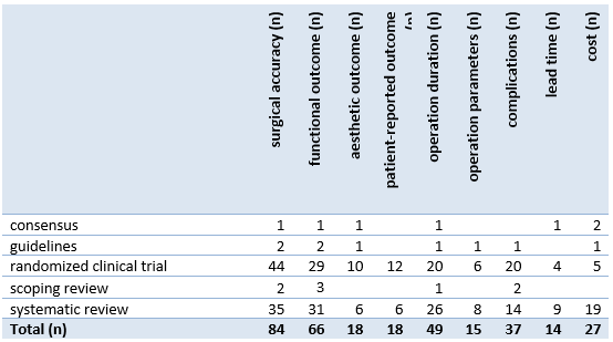
For further analysis of the found abstracts, the outcome measures were categorized; surgical accuracy, functional outcome, aesthetic outcome, patient-reported outcome, operation duration, operation parameters, complications, lead time and costs, as shown in Table 2.
Table 2. Overview outcome measure per publication
General oral and maxillofacial surgery
Fifteen systematic reviews and one consensus paper did not focus on a specific subspecialty but considered 3D printing in oral and maxillofacial surgery in general, or surgery with oral and maxillofacial as a subset (Choi, 2015; Bauermeister, 2016; Tack, 2016; Diment, 2017; Jacobs, 2017; Louvrier, 2017; Otero, 2017; Dion, 2018; Serrano, 2019; Ballard, 2020; Meglioli, 2020; Goodson, 2021; Ahmed, 2022; Meister, 2024; Tel, 2024; Kattimani, 2025; Sharaf, 2025).) Table 3 gives an overview of the 3D print types used in these publications of general oral and maxillofacial surgery.
Anatomical models were mentioned in 8/16 publications, mainly for preoperative planning, simulation, and intraoperative reference. Reduction in operation duration was specified as the main advantage of these models (Tack, 2016; Diment, 2017; Louvrier, 2017; Ballard, 2020; Meister, 2024),while increased cost was stated as a drawback (Tack, 2016; Jacobs, 2017; Louvrier, 2017; Ballard, 2020).
Patient-specific implants (PSIs) were also addressed in 8/16 publications (Bauermeister, 2016; Tack, 2016; Jacobs, 2017; Louvrier, 2017; Dion, 2018; Goodson, 2021; Ahmed, 2022; Kattimani, 2025). These studies reported overall favorable functional outcome and reduced operation duration for 3D-printed PSIs, but also an increase in cost. The consensus publication (Goodson (2021)) found overall consensus about the use of PSI in orbital reconstruction and oncologic reconstruction with fibula graft.
The findings for surgical guides (5/16) were used to transfer planned drill positions, screw positions, or osteotomies to the surgical field. Splints (2/16) were applied for repositioning of the jaw in orthognathic surgery, whereas bending guides (1/16) supported pre-contouring of fixation plates.
Table 3. Overview print type per publication (general oral and maxillofacial surgery)

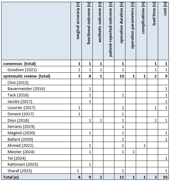
In addition to print type, the outcome measures of the publications have been analyzed, as shown in Table 4. Reporting most often addressed operation duration (10/16), functional outcomes (9/16), and cost (9/16). Most publications assessed a variety of 3D print types, hampering distinction between different types in the analysis of results.
Table 4. Overview outcome measure per publication (general oral and maxillofacial surgery)
Summary
Across oral and maxillofacial surgery, 3D printing is most commonly used for anatomical models, patient-specific implants, and surgical guides. Most studies, regardless of device, report similar patterns: predictable and (more) accurate translation of virtual plans to surgery, good functional outcome, and faster intraoperative execution. Direct cost are reported to be increased, although time savings may make up for it in cost effectiveness analysis.
Implantology
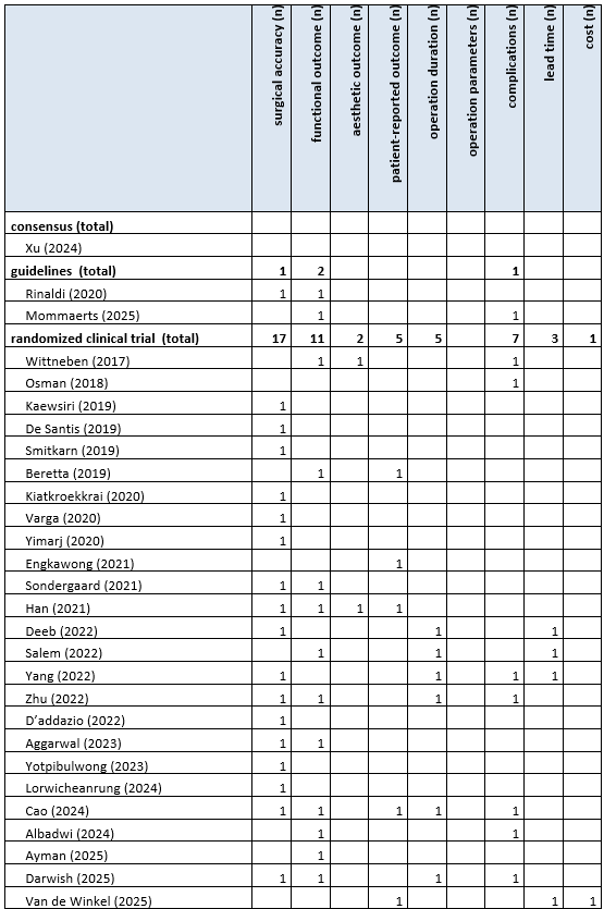
Across 42 implantology publications, 3D printing was applied for surgical guide fabrication, implant and abutment design, and planning models in alveolar, maxillary, and mandibular procedures (Fuster-Torres, 2009; Wittneben, 2017; Osman, 2018; Seo, 2018; Wismeijer, 2018; Beretta, 2019; De Santis, 2019; Kaewsiri, 2019; Smitkarn, 2019; Kiatkroekkrai, 2020; Rinaldi, 2020; Varga, 2020; Yimarj, 2020; Engkawong, 2021; Han, 2021; Søndergaard, 2021; Abu-Mostafa, 2022; Deeb, 2022; D’Addazio, 2022; Salem, 2022; Salinero, 2022; Yang, 2022; Zhu, 2022; Aggarwal, 2023; Balaguer-Martí, 2023; Marques-Guasch, 2023; Shi, 2023; Takács, 2023; Tanveer, 2023; Yotpibulwong, 2023; Albadwi, 2024; Cao, 2024; Lorwicheanrung, 2024; Xu, 2024; Ayman, 2025; Baecher, 2025; Darwish, 2025; Dipalma, 2025; Mommaerts, 2025; Sudhir, 2025; Van de Winkel, 2025). Reported outcomes most frequently included surgical accuracy (26/42), functional outcomes (19/42), and complications (12/42), with less focus on patient-reported (6/42), operation duration (5/42), and cost (4/42). Table 5 shows an overview of print types per publication about implantology. In Table 6 the outcome measures per publication are shown.
Table 5. Overview print type per publication (implantology)


Table 6. Overview outcome measure per publication (implantology)
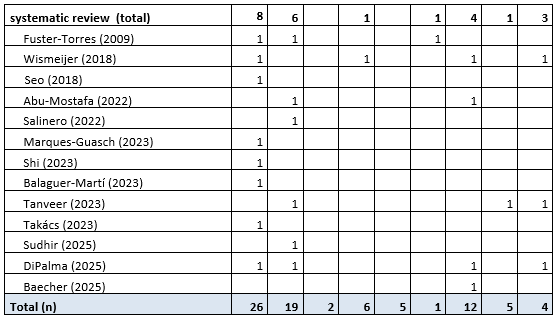
Surgical guides (28/42) were the most frequently used 3D-printed devices in implantology. Guides were designed to ensure precise implant positioning based on virtual planning. Studies comparing a guided approach to freehand implant placement found compelling evidence for superior accuracy for guided implant positioning (Fuster-Torres, 2009; Aggarwal, 2023; Marques-Guasch, 2023; Takács, 2023; Kaewsiri, 2019; Smitkarn, 2019; Kiatkroekkrai, 2020; Varga, 2020; Yimarj, 2020; Deeb, 2022; Seo, 2022; Yotpibulwong, 2023; Lorwicheanrung, 2024). Typical reported deviations of a guided approach were around 1 mm at the implant platform, around 1.5 mm at the apex and an angular deviation of roughly 3 degrees (Kaewsiri, 2019; Kiatkroekkrai, 2020; Yimarj, 2020; Yotpibulwong, 2023).). In studies where the 3D printed guides were compared to surgical navigation, no significant differences in positioning were found, a combined approach yielded improved accuracy over the individual approaches in two randomized controlled trials (Yotpibulwong,2023; Lorwicheanrung, 2024). Some studies also described reduced complications or operation duration with a guided approach.
Prosthetic components (6/42) covered a variety of custom restorations including crowns and dentures (Tanveer, 2023; Albadwi, 2024; Cao, 2024; Baecher, 2025; Van de Winkel, 2025). While the amount of evidence in each outcome category is limited 3D printed prosthetic components had favourable outcome in nearly all outcome categories. Patient-specific implants (PSIs) (5/42) were used in facial implantology or in case of individualized subperiostal implantology in the atrophic mandible or maxilla. Overall, functional outcome was reported to be positive in this limited amount of studies. 3D-printed abutments (3/40) were used for customized prosthetic connections. Functional outcome of this approach was appraised positively (Fuster-Torres, 2009; Wittneben, 2017; Beretta, 2019).
Summary
In implantology, 3D printing is most frequently used for surgical guides, where evidence for improved surgical accuracy with the use of 3D printed guides was reported. Prosthetic components, customized abutments, or patient-specific implants in complex cases with severe atrophy are much less frequently used. Patient-reported outcomes are only occasionally addressed, and cost-effectiveness data remain sparse. Overall, for 3D printing in implantology well-established advantages in surgical precision and functional outcome were reported.
Orthognathic surgery
Across 30 orthognathic surgery publications, 3D printing was used in single- and bimaxillary procedures for repositioning of mandible or maxilla in monomaxillary or bimaxillary surgery, with the help of splints, guides, or patient-specific implants (Hsui-Hsia, 2018; Van den Bempt, 2018; Hanafy, 2019; Schneider, 2019; Hanafy, 2020; Kraeima, 2020; Li, 2020; Chen, 2021; Figueiredo, 2021; Li, 2021; Liu, 2021; Cui, 2022; Goulart, 2022; Hamdy Mahmoud, 2022; Sánchez-Jáuregui, 2022; Alhabshi, 2023; Diaconu, 2023; Pellegrini, 2023; van der Wel, 2023; Lee, 2024; On, 2024; Straub, 2024; Abdelhamid, 2025; Bevini, 2025; Kobravi, 2025; Morsi, 2025; Nastri, 2025; Nguyen, 2025; Schrader, 2025; van der Wel, 2025). Reporting focus was almost always on surgical accuracy (25/30); in contrast, other outcome parameters were infrequently described (functional outcomes (7/30), operation duration (5/30), complications (3/30), cost (3/30), aesthetic outcomes (3/30), patient-reported outcomes (3/30), operative parameters (1/30)). Table 7 shows an overview of print types per publication about orthognathic surgery. In Table 8 the outcome measures per publication are shown.
Table 7. Overview print type per publication (orthognathic surgery)

Table 8. Overview outcome measure per publication (orthognathic surgery)
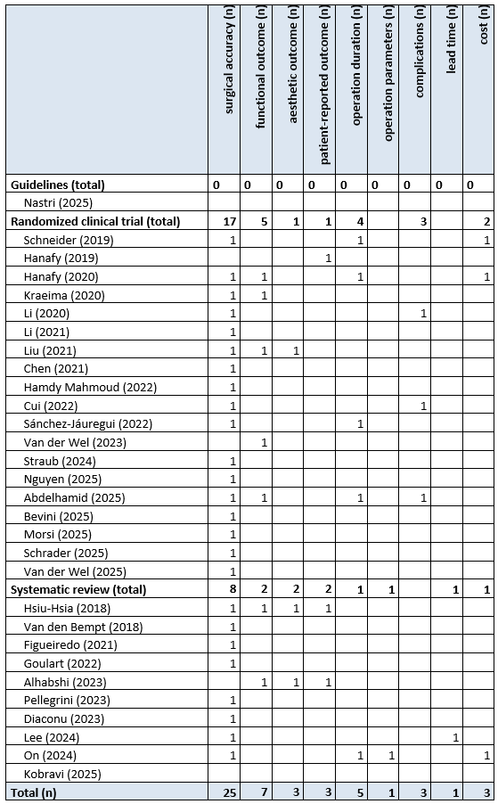
3D-printed splints (18/30) were the most frequently used 3D-printed device type in orthognathic surgery. Splints can be seen as positioning guides based on the dentition: splint contains dental impressions of the maxilla and mandible, where either one dental arch impression is in the planned position relative to the preoperative position of the other arch (intermediate splint; either maxilla-first or mandible-first depending on the repositioning order) or both are in the new position (final splint). Surgical accuracy is assessed in 17/18 studies, but splints were only compared to non-3D-printed splints in three publications; one publication favoured 3D printed splints (Schneider (2019)), one found no significant difference (Chen (2021)), and one had contradictory statements in the abstract (Nguyen (2025)). More often, the 3D printed splints were considered the control group for newer positioning approaches.
The use of patient-specific implants (PSIs) (12/30) can be considered one of these novel 3D printed tools. The approach may differ within publications: some studies only use patient-specific implants for the first repositioning (mainly maxilla-only), while others use PSIs in both maxilla and mandible repositioning. The accuracy of PSIs was compared to 3D printed wafers in seven PSI publications; (Dianocu, 2023; Kraeima, 2020; Chen, 2021; Li, 2021; Sánchez-Jáuregui, 2022; Van der Wel, 2025) concluded a significantly improved accuracy was obtained with PSI, while Van den Bempt (2018) was unclear on the topic. Functional outcomes were described only by Van der Wel (2023) as skeletally stable for both 3D printed approaches in one publication, while Kraeima (2020). mentioned a clinically relevant cutoff point for the use of PSI Surgery duration was the only parameter assessed outside of surgical accuracy and functional outcome by (Sánchez-Jáuregui (2022)).
Surgical guides (11/30) are used to transfer osteotomy lines and fixation positions in single- and bimaxillary procedures. While these are considered mandatory in the PSI approach, the overlap between both groups was only 50%, indicating severe underreporting in either one or both. Two publications described a combination with prebent osteosynthesis material (Li (2020) and Liu (2021)), of which at least one used it in combination with a 3D printed bending guide (Li (2020)). Cui (2022) described a different use case: 3D guided positioning of intermaxillary fixation screws, and found more accurate positioning with less complications than the conventional approach.
Summary
Across orthognathic surgery literature, focus was on accuracy. 3D printed devices were frequently compared against other 3D printed applications, demonstrating the extent of 3D printing in orthognathic surgery. Overall, 3D printing in orthognathic surgery is reported to support accurate plan transfer, but functional and patient-centered evidence remain sparse.
Craniomaxillofacial traumatology
21 publications reported the use of 3D printing in surgical intervention after craniomaxillofacial trauma (Prisman, 2014; Raisian, 2017; Engels, 2019; Azarmehr, 2020; Ramanathan, 2020; Zhao, 2021; Chepurnyi, 2022; Maher, 2022; Murray-Douglass, 2022; Truscott, 2022; Kotecha, 2023; Singh, 2023; Zhang, 2023; Amin, 2024; Committeri, 2024; Oley, 2024; Sintuwong, 2024; Valls-Esteve, 2024; Bertin, 2025; DiPalma, 2025; Helgers, 2025). The main focus within craniomaxillofacial trauma was on orbital reconstruction, which was (one of) the main topic(s) in two thirds of publications (Raisian, 2017; Engels, 2019; Azarmehr, 2020; Chepurnyi, 2022; Maher, 2022; Murray-Douglass, 2022; Kotecha, 2023; Singh, 2023; Zhang, 2023; Amin, 2024; Sintuwong, 2024; Bertin, 2025; DiPalma, 2025; Helgers, 2025); in contrast, a limited number of publications specified mandibular trauma (5/21) (Prisman, 2014; Ramanathan, 2020; Zhao, 2021; Truscott, 2022; Bertin, 2025) or midface trauma (2/21) (Committeri, 2024; Bertin, 2025) as possible indication for 3D printing. Reporting focused on functional outcomes (14/21) and surgical accuracy (13/21), and operation duration (10/21), and complications. In contrast, cost (4/21), aesthetic outcomes (2/21), and patient-reported outcomes (2/21) were seldom assessed. Table 9 shows an overview of print types per publication about craniomaxillofacial traumatology. In Table 10 the outcome measures per publication are shown.
Table 9. Overview print type per publication (craniomaxillofacial traumatology)

Table 10. Overview outcome measure per publication (craniomaxillofacial traumatology)
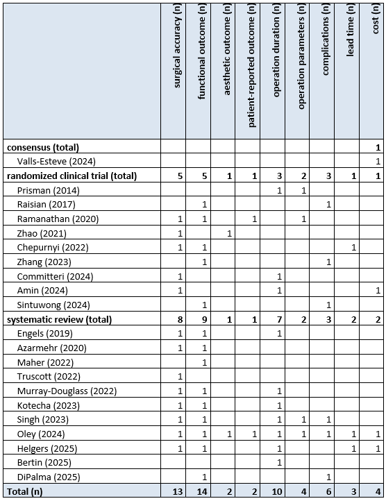
Bending guides (10/21) were the most frequently used 3D-printed device type in craniomaxillofacial trauma. These were predominantly applied in orbital wall/floor fracture repair or mandibular reconstruction. The guides enabled preoperative plate contouring to match the patient-specific bony anatomy. Reduction of surgical duration – likely because of time saved on intraoperative bending – was reported in seven publications (Engels, 2019; Singh, 2023; Helgers, 2025; Bertin, 2025; Prisman, 2014; Amin, 2024; Committeri, 2024). Studies on orbital reconstruction reported improved surgical accuracy, volume restoration of the orbit, and functional outcome in terms of diplopia and enophthalmos with guide-bended implants (Engels, 2019; Maher, 2022; Singh, 2023; Helgers, 2025; Raisian, 2017; Zhang, 2023; Amin, 2024). Complication reporting was limited (3/10) and no hard conclusions could be drawn from pooling the information.
Patient-specific implants (PSIs) (7/21) present an alternative for shaping osteosynthesis material: instead of bending on a model, the plate is directly 3D printed. Its use was again mainly described in orbital reconstruction: while two publications provided overarching data on its use in craniomaxillofacial trauma (Oley, 2024; DiPalma, 2025), the other publications specifically delved into orbital reconstruction. Orbital reconstruction with PSI achieved accurate reconstruction and adequate restoration of orbital contour and volume (Azarmehr, 2020; Kotecha, 2023; Helgers, 2025; Chepurnyi, 2022). Functional outcomes in terms of enophthalmos and diplopia were frequently reported as positive, but the two publications comparing it to other reconstruction could not find significant support for the use of PSIs in terms of functional outcome (Maher, 2022; Kotecha, 2023).
3D printing of anatomical models (3/21); surgical guides (3/21); splints (2/21); or prosthetics (2/21) was infrequently reported, mainly for specific indications (splints for mandibular fractures, prosthetics for orbital implants after enucleation). No conclusions on efficacy of these 3D printed models could be drawn from the small sample of publications.
Summary
In trauma surgery, 3D-printed bending guides and patient-specific implants represent the most frequently studied applications, primarily for orbital and mandibular reconstruction. Both are primarily used in orbital reconstruction, to either prebend the orbital reconstruction plate or to directly 3D print it. Evidence is primarily reporting increased accuracy through 3D printing, while the effect on other outcome parameters is not as clear.
Head and neck oncology
Across 21 head and neck oncology publications, oncologic resection and reconstruction of maxillary and mandibular tumors were most frequently given as the indication (De Farias (2014); Prisman (2014); Rodby (2014); Tarsitano (2015); Lin (2018); van Baar (2018); Ong (2019); Gupta (2021); Padilla (2021); van Baar (2021); Al-Sabahi (2022); Alwadeai (2022); Salinero (2022); Truscott (2022); Gernandt (2023); Juanita (2023); Alwadeai (2024); Chen (2024); Malzoni (2024); Serioli (2024); Srivastava (2024)). Table 11 shows an overview of print types per publication about head and neck oncology. In Table 12 the outcome measures per publication are shown.
Table 11. Overview print type per publication (head and neck oncology)

Table 12. Overview outcome measure per publication (head and neck oncology)
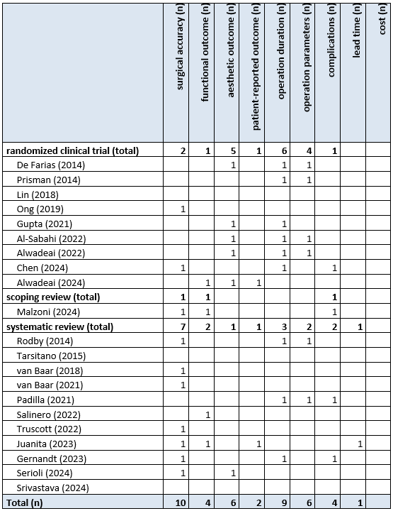
Reporting addressed surgical accuracy in more than half of the publications (11/21), followed by operation duration (9/21); a small amount reported complications (5/21) or aesthetic (6/21), functional (4/21), or patient-reported outcome (3/21). In over one third of publications (8/21), the exact device was not specified. These publications generally describe accuracy improvement and reduced operation time with 3D printing and better aesthetic outcome, but attributing these results to one of the printing categories below is infeasible. It seems also reasonable to assume that some 3D printed devices were used in conjunction with others (i.e. osteotomy guides to resect the mandible and fibula and a bending guide to mold the fixation plate to the neomandible), although this was again not always specified.
Decreased operation duration – with significantly lower ischaemia time – were also stated as study findings in 3/6 publications on surgical guides (Gernandt (2023); Al-Sabahi (2022); Alwadeai (2022)). Reconstruction aesthetics were also found to be significantly improved through the use of 3D printing in three randomized controlled trials (Al-Sabahi (2022); Alwadeai (2022); Alwadeai (2024)). Bending guides (3/21) supported preoperative contouring of fixation plates to patient-specific geometry. The use of patient-specific implants (PSIs) (2/21), anatomical models (2/21), obturators (1/21), prosthetic (custom) devices (1/21), or scaffolds (1/21) was found to be very limited in the publications included.
Summary
Oncologic reconstruction is well represented in 3D printed craniomaxillofacial indications. Overall improvement of accuracy and operation duration, combined with decrease in operation duration (and ischemia time) are the main reported rationales behind the use of 3D printed devices. In publications that did specify type of 3D print, surgical guides were the most frequently mentioned. The lack of specification within the abstracts hampers subset analysis of different 3D printing types.
Bone augmentation
Across 13 bone augmentation publications, 3D printing was applied for preoperative design and fabrication of scaffolds, custom implants, and surgical models to support alveolar maxillary and mandibular augmentation (Osman (2018); Maroulakos (2019); Abu-Mostafa (2022); Zhou (2022); Zhu (2022); Mohaghegh (2023); Cucchi (2024); Cucchi (2024); Giragosyan (2024); Ayman (2025); Cucchi (2025); Elrefaei (2025); Helal (2025)). Reported outcomes most frequently included functional outcomes (9/13) and complications (8/13), with limited data on other parameters. Table 13 shows an overview of print types per publication about bone augmentation. In Table 14 the outcome measures per publication are shown.
Table 13. Overview print type per publication (bone augmentation)

Table 14. Overview outcome measure per publication (bone augmentation)
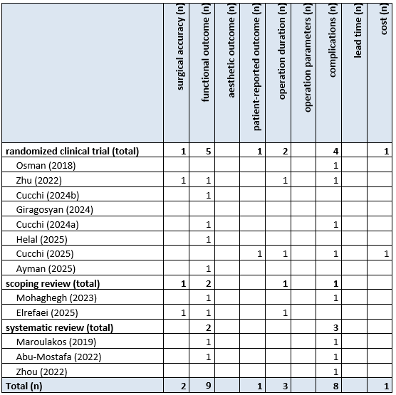
3D-printed scaffolds (10/13) were the most frequently reported application in bone augmentation. Scaffolds were typically fabricated for alveolar or sinus floor augmentation, serving as osteoconductive matrices or space-maintaining devices. Titanium was the most reported material (7/13) (Abu-Mostafa (2022); Cucchi (2024); Cucchi (2024); Cucchi (2025); Elrefaei (2025); Maroulakos (2019); Zhou (2022)); other materials (PEEK and hydroxyapatite) were reported only once (Elrefaei (2025); Maroulakos (2019)). Studies described consistent shape fidelity, good fit to the defect, and satisfactory new bone formation over time (Zhu (2022); Cucchi (2024); Elrefaei (2025); Abu-Mostafa (2022); Helal (2025); Maroulakos (2019); Ayman (2025)). Quantitative volumetric results showed bone gain comparable or superior to conventional grafting in several cases. Complications were deemed low overall, with some studies reporting a lower complication rate for a 3D printed approach whereas others did not.
Surgical guides (3/13) were applied to guide the positioning of grafts, scaffolds, or fixation devices during augmentation. In this limited sample, guides were said to ensure easy positioning of augmentation materials, reduce manual intraoperative adjustment, and improve accuracy (Osman (2018); Zhu (2022); Ayman (2025)). Bending guides (3/13) were compared to 3D printed scaffolds in three randomized clinical trials, with no significant differences reported between all outcomes (Abu-Mostafa (2022); Cucchi (2024); Cucchi (2025)).
Summary
Across bone augmentation studies, 3D printing was most frequently used to fabricate a scaffold. It was reported that it enables personalized reconstruction and technical precision, with accurate defect matching and good regenerative potential reported. Reported evidence primarily documents functional outcome in terms of bone gain, where 3D printed scaffolds ensure bone formation.
Other applications in oral and maxillofacial surgery
Twenty-two publications had indications that were found five times or less (Ritschl, 2016; Kim, 2018; Hou, 2020; Yang, 2020; Cao, 2021; Chiesa-Estomba, 2021; Hosameldin, 2021; Kerbrat, 2021; Kumar, 2021; Sembronio, 2021; Tanveer, 2021; Tanveer, 2021; Olate, 2023; Abdelwahed, 2024; Clegg, 2024; Goetze, 2024; Gursky, 2024; Ma, 2024; Chowdhury, 2025; Di Cosmo, 2025; Rana, 2025; Tachizawa, 2025). Cranial reconstruction was the shared largest indication in the ‘other’ group (5/22) (Yang, 2020; Hosameldin, 2021; Abdelwahed, 2024; Di Cosmo, 2025; Rana, 2025). Table 15 shows an overview of print types per publication about other applications in oral and maxillofacial surgery. In Table 16 the outcome measures per publication are shown.
Table 15. Overview print type per publication (other applications)

Table 16. Overview print type other applications

In all studies, patient-specific implants (PSIs) in PEEK, titanium, or ultra-high-molecular weight polyethylene (UHMWPE) were used as a 3D device (3D printing or milling was not always specified). In all publications, customized implants were found to be superior to conventional implants for every outcome parameter. Nose reconstruction (5/22) was described as often as cranial reconstruction (Ritschl, 2016; Cao, 2021; Chiesa-Estomba, 2021; Tanveer, 2021; Chowdhury, 2025). Scaffolds were predominantly 3D printed (Cao, 2021; Chiesa-Estomba, 2021; Chowdhury, 2025), but since two of these (Cao, 2021 and Chiesa-Estomba, 2021) did not specify any outcome parameter, any conclusion is infeasible.
Four studies had genioplasty as their main indication (Kim, 2018; Ma, 2024; Gursky, 2024; Tachizawa, 2025); the publication with the most patients (Gursky (2024)) reviewed genioplasty in light of facial feminization surgery. In three publications, high patient satisfaction through 3D printed devices was reported (Kim, 2018; Ma, 2024; Gursky, 2024). In TMJ reconstruction (3/22) (Kumar, 2021; Sembronio, 2021; Olate, 2023), focus was again on PSIs (2/3; Kumar (2021) did not specify printing type). Improved functional outcome was reported in the short term (Kumar (2021) and Olate (2023)), while long term results remain to be seen.
Only one publication was found for orbital prosthesis (Tanveer, 2021), pediatric craniomaxillofacial surgery (Clegg 2024), auto transplantation (Hou, 2020), obstructive sleep apnea (Kerbrat, 2021) or on Medical Device Regulations in oral and maxillofacial surgery (Goetze, 2024).
No systematic review of the literature has been performed as it is not conceivable that a research design can be used to answer the main question of the module. Although no systematic review based on a PICO, including a GRADE judgement, could be performed, a review of available literature was performed to answer the following questions:
- What kind of 3D print clinical applications or 3D printed tools are described in literature regarding oral and maxillofacial surgery?
- per indication.
- Which studies describe the added value of 3D printing in oral and maxillofacial surgery?
- per indication.
Methods
A systematic literature search was performed by a medical information specialist using the following bibliographic databases: Embase.com and Ovid/Medline. Both databases were searched from 2005 to the 18th of September 2025 for guidelines, systematic reviews, RCTs, observational studies and reviews. Systematic searches were completed using a combination of controlled vocabulary/subject headings (e.g., Emtree-terms, MeSH) wherever they were available and natural language keywords. The overall search strategy was derived from six primary search concepts: (1) 3D printing; (2) anatomic model; (3) surgical guide; (4) mold; (5) patient specific implants; (6) oral and maxillofacial surgery. Duplicates were removed using EndNote software. After deduplication and removing of retracted articles a total of 2085 records were imported for title/abstract screening.
Because of the large number of studies found, only randomized controlled trials, guidelines, expert consensus, or systematic reviews were considered for inclusion. Exclusion criteria were:
- No abstract available.
- No description of the use of 3D printing in title or abstract.
- Not related to oral and maxillofacial surgery1.
- Not related to patient care.
- In-vitro, ex-vivo, or animal studies.
- Non-English language.
Ad1: publications on dentistry with no oral and maxillofacial surgeons in the author list were also removed, since these interventions were expected to take place in dental clinics rather than hospitals.
The method for identification, screening and inclusion of literature is schematically shown in Figure 1. It should be noted that the literature analysis was performed using only the information available in the abstract.
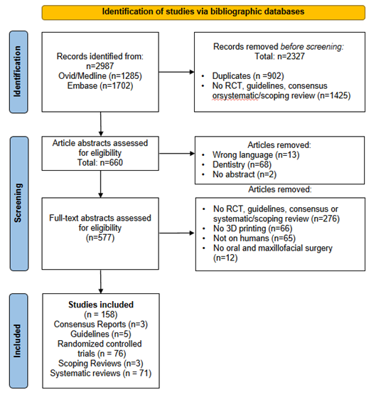
Figure 1. Flow diagram of the identification, screening and selection process for the abstracts
- Abdelhamid, Abdelkareem M. and Hassan, Atef M. and El-Mohandes, Wael A. Accuracy Assessment of Customized Titanium Plates Compared to 3D-Printed Splints in Le Fort I Osteotomy: A Randomized Clinical Trial Evaluating Clinical and Radiographic Outcomes. Cureus. 2025; 17 (5):e84151.
- Abdelwahed, M. S. and Ali, S. and Abdelwahed, A. S. K. and Aziz, M. M. and Bassiouny, M. S. and Ahmed, M. S. Cranioplasty using patient specific implants Polyether ether ketone versus ultra-high molecular weight polyethylene: A prospective study. Journal of Cranio-Maxillofacial Surgery. 2024; 52 (11):1299-1310.
- Abu-Mostafa, N. A. and Alotaibi, Y. N. and Alkahtani, R. N. and Almutairi, F. K. and Alfaifi, A. A. and Alshahrani, O. D. The Outcomes of Vertical Alveolar Bone Augmentation by Guided Bone Regeneration with Titanium Mesh: A Systematic Review. The journal of contemporary dental practice. 2022; 23 (12):1280-1288.
- Aggarwal, S. and Aggarwal, S. and Goswami, R. and Mowar, A. and Tomar, N. and Saxena, D. An in vivo study to assess and compare the angular, linear, and depth deviation as well as the difference in bone density of implants placed using computer-aided design/computer-aided manufacturing fabricated three-dimensional guides versus the implants placed using bone pen kit in maxillary and .... Journal of Indian Prosthodontic Society. 2023; 23 (3):266-276.
- Ahmed, K. S. and Ibad, H. and Suchal, Z. A. and Gosain, A. K. Implementation of 3D Printing and Computer-Aided Design and Manufacturing (CAD/CAM) in Craniofacial Reconstruction. The Journal of craniofacial surgery. 2022; 33 (6):1714-1719.
- Al-Sabahi, Mohammed Esmail and Jamali, Omer Mohammed and Shindy, Mostafa Ibrahim and Moussa, Basma Gamal and Amin, Ayman Abdel-Wahab and Zedan, Mohamed Hamdallah Aesthetic Reconstruction of Onco-surgical Mandibular Defects Using Free Fibular Flap with and without CAD/CAM Customized Osteotomy Guide: A Randomized Controlled Clinical Trial. BMC cancer. 2022; 22 (1):1252.
- Alhabshi, Manaf O. and Aldhohayan, Haya and BaEissa, Olla S. and Al Shehri, Manar S. and Alotaibi, Nawaf M. and Almubarak, Sarah K. and Al Ahmari, Abdulrahman A. and Khan, Hayithm A. and Alowaimer, Hesham A. Role of Three-Dimensional Printing in Treatment Planning for Orthognathic Surgery: A Systematic Review. Cureus. 2023; 15 (10):e47979.
- Alwadeai, M. and Al-aroomy, L. and Amin, A. and Shindy, M. and Zedan, M. and Baz, S. Virtual Surgical Guidance Improves Quality of Life Following Scapular Free-Flap Reconstruction of Maxillary Defects. Journal of Oral and Maxillofacial Surgery. 2024; 82 (5):600-609.
- Alwadeai, Mohamed Salah and Al-Aroomy, Leena Ali and Shindy, Mostafa Ibrahim and Amin, Ayman Abdel-Wahab and Zedan, Mohamed Hamdallah Aesthetic reconstruction of onco-surgical maxillary defects using free scapular flap with and without CAD/CAM customized osteotomy guide. BMC surgery. 2022; 22 (1):362.
- Amin, D. and Nguyen, N. and Manhan, A. J. and Kim, J. H. and Roser, S. M. and Bouloux, G. F. Does a Point-of-Care 3-Dimensional Printer Result in a Decreased Length of Surgery for Orbital Fractures?. Journal of Oral and Maxillofacial Surgery. 2024; 82 (10):1275-1284.
- Ayman, D. and Shawky, M. and Aly, L. A. A. and Mounir, M. and Zekry, A. K. A. Bone gain and accuracy assessment of computer-guided workflow for horizontal augmentation of atrophic anterior maxilla with symphyseal cortical plates: a randomized controlled trial. BMC oral health. 2025; 25 (1):1039.
- Azarmehr, I. and Stokbro, K. and Bell, R. B. and Thygesen, T. Contemporary Techniques in Orbital Reconstruction: A Review of the Literature and Report of a Case Combining Surgical Navigation, Computer-Aided Surgical Simulation, and a Patient-Specific Implant. Journal of Oral and Maxillofacial Surgery. 2020; 78 (4):594-609.
- Baecher, Helena and Scheiflinger, Alexandra and Remy, Katya and Straub, Niklas and Maheta, Bhagvat and Sherwani, Khalil and Deniz, Can and Knoedler, Samuel and Safi, Ali-Farid and Kauke-Navarro, Martin and Heiland, Max and Knoedler, Leonard From Novel Facial Measurements to Facial Implantology: A Systematic Review. JPRAS open. 2025; 43:1-17.
- Balaguer-Martí, J. C. and Canet-López, Á and Peñarrocha-Diago, M. and Romeo-Rubio, M. and Peñarrocha-Diago, M. and García-Mira, B. Influence of Splint Support on the Precision of Static Totally Guided Dental Implant Surgery: A Systematic Review and Network Meta-analysis. International Journal of Oral and Maxillofacial Implants. 2023; 38 (1):157-168.
- Ballard, D. H. and Mills, P. and Duszak, R. and Weisman, J. A. and Rybicki, F. J. and Woodard, P. K. Medical 3D Printing Cost-Savings in Orthopedic and Maxillofacial Surgery: Cost Analysis of Operating Room Time Saved with 3D Printed Anatomic Models and Surgical Guides. Academic Radiology. 2020; 27 (8):1103-1113.
- Bauermeister, A. J. and Zuriarrain, A. and Newman, M. I. Three-Dimensional Printing in Plastic and Reconstructive Surgery: A Systematic Review. Annals of plastic surgery. 2016; 77 (5):569-576.
- Bevini, M. and Zavalloni, G. and Federico, L. and Centra, M. and Gulotta, C. and Mirabile, F. M. C. and Cercenelli, L. and Incerti Parenti, S. and Marcelli, E. and Tarsitano, A. and Badiali, G. Single-jaw patient-specific implants in bimaxillary orthognathic surgery: Randomized cross-controlled comparison between maxilla-guided and mandible-guided approach. Journal of Cranio-Maxillofacial Surgery. 2025; 53 (5):632-641.
- Cao, Y. and Hu, M. and Zhang, J. and Yu, L. and Gao, Y. and Ding, Z. and Ma, H. and Zhu, F. Analysis of the efficacy of two kinds of loss restoration of posterior teeth using 3D printing temporary crown during the second phase of implant surgery. Medicine (United States). 2024; 103 (48):e40460.
- Chen, H. and Bi, R. and Hu, Z. and Chen, J. and Jiang, N. and Wu, G. and Li, Y. and Luo, E. and Zhu, S. Comparison of three different types of splints and templates for maxilla repositioning in bimaxillary orthognathic surgery: a randomized controlled trial. International Journal of Oral and Maxillofacial Surgery. 2021; 50 (5):635-642.
- Chen, T. and Chan, H. H. L. and de Almeida, J. and Goldstein, D. P. and Gilbert, R. W. and Yao, C. M. K. L. and Irish, J. C. and Davies, J. C. A 3D Analysis of Plating Strategies in Mandibular Reconstruction: A Randomized Control Pilot Study. Laryngoscope. 2024; 134 (5):2182-2186.
- Chepurnyi, Y. and Zhukovtseva, O. and Kopchak, A. and Kanura, O. Clinical application of automated virtual orbital reconstruction for orbital fracture management with patient-specific implants: A prospective comparative study. Journal of Cranio-Maxillofacial Surgery. 2022; 50 (9):686-691.
- Chiesa-Estomba, C. M. and Aiastui, A. and González-Fernández, I. and Hernáez-Moya, R. and Rodiño, C. and Delgado, A. and Garces, J. P. and Paredes-Puente, J. and Aldazabal, J. and Altuna, X. and Izeta, A. Three-Dimensional Bioprinting Scaffolding for Nasal Cartilage Defects: A Systematic Review. Tissue Engineering and Regenerative Medicine. 2021; 18 (3):343-353.
- Choi, J. W. and Kim, N. Clinical application of three-dimensional printing technology in craniofacial plastic surgery. Archives of Plastic Surgery. 2015; 42 (3):267-277.
- Committeri, U. and Magliulo, R. and Carraturo, E. and Arena, A. and Abbate, V. and Salzano, G. and Troise, S. and Barone, S. and Germano, C. and Vaira, L. A. and Giovacchini, F. and Cataldo, R. and Grassia, M. G. and Califano, L. and Piombino, P. Virtual surgical planning in tripod zygomatico-maxillary complex fractures: A prospective comparison between two different strategies. Journal of Cranio-Maxillofacial Surgery. 2024; 52 (12):1497-1504.
- Cucchi, A. and Bettini, S. and Tedeschi, L. and Franceschi, D. and Urban, I. and Fiorino, A. and Corinaldesi, G. Operative Times, Costs and Patient-Related Outcome Measures in Vertical Ridge Augmentation With Customised Reinforced PTFE Mesh Versus CAD/CAM Titanium Mesh: Secondary Analysis of a Randomised Clinical Trial. Journal of Clinical Periodontology. 2025; 52 (7):971-982.
- Cucchi, A. and Bettini, S. and Tedeschi, L. and Urban, I. and Franceschi, D. and Fiorino, A. and Corinaldesi, G. Complication, vertical bone gain, volumetric changes after vertical ridge augmentation using customized reinforced PTFE mesh or Ti-mesh. A non-inferiority randomized clinical trial. Clinical oral implants research. 2024; 35 (12):1616-1639.
- Cucchi, A. and Marchiori, G. and Sartori, M. and Fini, M. and Fiorino, A. and Donati, R. and Corinaldesi, G. and Maglio, M. A 3D micro-CT assessment of composition and structure of bone tissue after vertical and horizontal alveolar ridge augmentation using CAD/CAM-customized titanium mesh. Clinical oral implants research. 2024; 35 (12):1546-1559.
- Cui, Ming-Xue and Xiao, Le-Cun and Yue, Jin and Xue, Ling-Fa and Xiao, Wen-Lin Effect of a digital guide on the positional accuracy of intermaxillary fixation screw implantation in orthognathic surgery. Journal of plastic, reconstructive & aesthetic surgery : JPRAS. 2022; 75 (7):e15-e22.
- Darwish, Saad A. and El-Mohandes, Wael A. and Abd Rabbo, Bahaa El-Din Clinical and Radiographic Assessment of Milled Versus 3D-Printed Patient-Specific Subperiosteal Implants for Atrophic Mandibular Ridges: A Randomized Clinical Trial. Cureus. 2025; 17 (3):e80326.
- De Farias, T. P. and Dias, F. L. and Galvao, M. S. and Boasquevisque, E. and Pastl, A. C. and Sousa, B. A. Use of prototyping in preoperative planning for patients with head and neck tumors. Head and Neck. 2014; 36 (12):1773-1782.
- Deeb, J. G. and Kanduti, D. and Skrjanc, L. and Carrico, C. K. and Deeb, G. Comparison of Accuracy and Time for Four Implant Placement Techniques Supporting Fixed-Partial Denture. The Journal of oral implantology. 2022; 48 (6):562-572.
- Di Cosmo, L. and Pellicanò, F. and Choueiri, J. E. and Schifino, E. and Stefini, R. and Cannizzaro, D. Meta-analyses of the surgical outcomes using personalized 3D-printed titanium and PEEK vs. standard implants in cranial reconstruction in patients undergoing craniectomy. Neurosurgical Review. 2025;48 (1):312.
- Diment, L. E. and Thompson, M. S. and Bergmann, J. H. M. Clinical efficacy and effectiveness of 3D printing: A systematic review. BMJ Open. 2017; 7 (12):e016891.
- DiPalma, A. D. and Zia, H. and Goodwin, B. and Al-Shehab, U. and Bharadia, A. and Goldfarb, J. Efficacy of absorbable vs. non-absorbable patient-specific, 3D-printed implants for the treatment of facial bone fractures: a systematic review and meta-analysis. Oral and maxillofacial surgery. 2025; 29 (1):30.
- Dipalma, G. and Inchingolo, A. M. and Trilli, I. and Di Noia, A. and De Vecchio, G. and Palermo, A. and Inchingolo, F. and Inchingolo, A. D. Accuracy of the surgical template used in the placement of implants and orthodontic miniscrews. BMC oral health. 2025; 25 (1):999.
- Engels, T. and Waskiewicz, K. and Glineur, R. and Oth, O. Evaluation of the preoperative pre-bending of titanium mesh on a model obtained by 3D printing for treatment of orbital floor fractures: A systematic review of the literature and description of a new protocol. Revue Medicale de Bruxelles. 2019; 40 (6):502-508.
- Engkawong, S. and Mattheos, N. and Pisarnturakit, P. P. and Pimkhaokham, A. and Subbalekha, K. Comparing patient-reported outcomes and experiences among static, dynamic computer-aided, and conventional freehand dental implant placement: A randomized clinical trial. Clinical implant dentistry and related research. 2021; 23 (5):660-670.
- Figueiredo, C. E. and Paranhos, L. R. and da Silva, R. P. and Herval, ÁM and Blumenberg, C. and Zanetta-Barbosa, D. Accuracy of orthognathic surgery with customized titanium plates-Systematic review. Journal of stomatology, oral and maxillofacial surgery. 2021; 122 (1):88-97.
- Fuster-Torres, Ma Á and Albalat-Estela, S. and Alcañiz-Raya, M. and Peñarrocha-Diago, M. CAD / CAM dental systems in implant dentistry: Update. Medicina Oral, Patologia Oral y Cirugia Bucal. 2009; 14 (3):E141-E145.
- Gernandt, S. and Tomasella, O. and Scolozzi, P. and Fenelon, M. Contribution of 3D printing for the surgical management of jaws cysts and benign tumors: A systematic review of the literature. Journal of stomatology, oral and maxillofacial surgery. 2023; 124 (4):101433.
- Giragosyan, K. and Chenchev, I. and Ivanova, V. Linear bone gain and healing complication rate comparative outcomes following ridge augmentation with custom 3D printed titanium mesh vs Ti-reinforced dPTFE. A randomized clinical trial. Folia medica. 2024; 66 (4):505-514.
- Goetze, Elisabeth and Zeller, Alexander- N. and Pabst, Andreas Approaching 3D printing in oral and maxillofacial surgery - suggestions for structured clinical standards. Oral and maxillofacial surgery. 2024; 28 (2):795-802.
- Goodson, A. M. C. and Parmar, S. and Ganesh, S. and Zakai, D. and Shafi, A. and Wicks, C. and O'Connor, R. and Yeung, E. and Khalid, F. and Tahim, A. and Gowrishankar, S. and Hills, A. and Williams, E. M. Printed titanium implants in UK craniomaxillofacial surgery. Part II: perceived performance (outcomes, logistics, and costs). British Journal of Oral and Maxillofacial Surgery. 2021; 59 (3):320-328.
- Goulart, M. E. and Biegelmeyer, T. C. and Moreira-Souza, L. and Adami, C. R. and Deon, F. and Flores, I. L. and Gamba, T. O. What is the accuracy of the surgical guide in the planning of orthognathic surgeries? A systematic review. Medicina oral, patologia oral y cirugia bucal. 2022; 27 (2):e125-e134.
- Gupta, S. and Goil, P. Formulating an Easy, Affordable, and Reproducible Method for Virtual Planning and 3D Reconstruction: A State Institution's Approach for Mandibular Reconstruction. Annals of plastic surgery. 2021; 87 (1):65-72.
- Hamdy Mahmoud, M. and Ismail Elfaramawi, T. Maxillary stability in patients with skeletal class III malocclusion treated by bimaxillary orthognathic surgery: comparison of mandible-first and maxilla-first approaches in a randomised controlled study. British Journal of Oral and Maxillofacial Surgery. 2022; 60 (6):761-766.
- Hanafy, M. and Akoush, Y. and Abou-ElFetouh, A. and Mounir, R. M. Precision of orthognathic digital plan transfer using patient-specific cutting guides and osteosynthesis versus mixed analogue-digitally planned surgery: a randomized controlled clinical trial. International journal of oral and maxillofacial surgery. 2020; 49 (1):62-68.
- Hanafy, Mahmoud and Abou-Elfetouh, Adel and Mounir, Ragia M. Quality of life after different approaches of orthognathic surgery: a randomized controlled study. Minerva stomatologica. 2019; 68 (3):112-117.
- Helal, M. H. and Sheta, M. S. and Alsherif, A. A. and Hassan, M. A. and Aboushelib, M. N. and Ghouraba, R. F. The Effectiveness of Hyaluronic Acid on Prefabricated CAD CAM Bone Blocks for Ridge Augmentation: A Split Mouth Study. Clinical implant dentistry and related research. 2025; 27 (2):e70035.
- Hou, R. and Hui, X. Y. and Xu, G. J. and Yang, X. and Wang, H. G. and Xu, N. and Liu, Y. L. and Zhu, M. H. and Zhou, H. Z. Clinical observation of three-dimensional printing donor tooth model in peri-operative period of autotransplantation of tooth. Zhonghua kou qiang yi xue za zhi = Zhonghua kouqiang yixue zazhi = Chinese journal of stomatology. 2020; 55 (9):647-653.
- Jacobs, C. A. and Lin, A. Y. A New Classification of Three-Dimensional Printing Technologies: Systematic Review of Three-Dimensional Printing for Patient-Specific Craniomaxillofacial Surgery. Plastic and reconstructive surgery. 2017; 139 (5):1211-1220.
- Juanita, M. and Raodah and Inriany, N. and Endang, F. and Affandhy, S. and Alfian, R. and Habar, I. D. Rehabilitation of Maxillectomy Defect with Palatal Obturator Using Digital Technology: A Review. Journal of Population Therapeutics and Clinical Pharmacology. 2023; 30 (9):e150-e160.
- Kaewsiri, D. and Panmekiate, S. and Subbalekha, K. and Mattheos, N. and Pimkhaokham, A. The accuracy of static vs. dynamic computer-assisted implant surgery in single tooth space: A randomized controlled trial. Clinical oral implants research. 2019; 30 (6):505-514.
- Kattimani, Vivekanand and Sreeram, Roopa Rani and Panga, Gnana Sarita Kumari and Vasamsetti, Divya Bhavani and Tiwari, Rahul Patient-Specific Implants in Maxillo-Facial Reconstruction: Current Practices and Way Forward-An Overview of Systematic Reviews. Journal of maxillofacial and oral surgery. 2025; 24 (4) :1075-1089.
- Kerbrat, A. and Vinuesa, O. and Lavergne, F. and Aversenq, E. and Graml, A. and Kerbrat, J. B. and Trost, O. and Goudot, P. Clinical impact of two types of mandibular retention devices - A CAD/CAM design and a traditional design - On upper airway volume in obstructive sleep apnea patients. Journal of stomatology, oral and maxillofacial surgery. 2021; 122 (4):361-366.
- Kiatkroekkrai, P. and Takolpuckdee, C. and Subbalekha, K. and Mattheos, N. and Pimkhaokham, A. Accuracy of implant position when placed using static computer-assisted implant surgical guides manufactured with two different optical scanning techniques: a randomized clinical trial. International Journal of Oral and Maxillofacial Surgery. 2020; 49 (3):377-383.
- Kim, S. C. and Kwon, J. G. and Jeong, W. S. and Na, D. and Choi, J. W. One-Piece Mandibuloplasty Compared to Conventional Mandibuloplasty With Narrowing Genioplasty. The Journal of craniofacial surgery. 2018; 29 (5) :1161-1168.
- Kobravi, Sepehr and Jafari, Aida and Lotfalizadeh, Mohammadhassan and Azimi, Abolfazl Digital Innovations in Orthognathic Surgery: A Systematic Review of Virtual Surgical Planning, Digital Transfer, and Conventional Model Surgery. Orthodontics & craniofacial research. 2025; 28 (5):783-798.
- Kotecha, S. and Ferro, A. and Harrison, P. and Fan, K. Orbital reconstruction: a systematic review and meta-analysis evaluating the role of patient-specific implants. Oral and maxillofacial surgery. 2023; 27 (2):213-226.
- Kraeima, J. and Schepers, R. H. and Spijkervet, F. K. L. and Maal, T. J. J. and Baan, F. and Witjes, M. J. H. and Jansma, J. Splintless surgery using patient-specific osteosynthesis in Le Fort I osteotomies: a randomized controlled multi-centre trial. International Journal of Oral and Maxillofacial Surgery. 2020; 49 (4):454-460.
- Kumar, S. and Khanna, V. and Singh, B. P. and Mehrotra, D. and Patil, R. K. Impact of technology in temporomandibular joint reconstruction surgeries: A systematic review. Journal of Plastic, Reconstructive and Aesthetic Surgery. 2021; 74 (6):1331-1345.
- Lee, Yoon-Jo and Oh, Ji-Hyeon and Kim, Seong-Gon Virtual surgical plan with custom surgical guide for orthognathic surgery: systematic review and meta-analysis. Maxillofacial plastic and reconstructive surgery. 2024; 46 (1):39.
- Li, B. and Wei, H. and Jiang, T. and Qian, Y. and Zhang, T. and Yu, H. and Zhang, L. and Wang, X. Randomized Clinical Trial of the Accuracy of Patient-Specific Implants versus CAD/CAM Splints in Orthognathic Surgery. Plastic and reconstructive surgery. 2021; 148 (5):1101-1110.
- Li, K. and Li, J. and Du, W. and Xu, C. and Ye, B. and Luo, E. Computer-Aided Design and Manufacturing Cutting and Drilling Guides with Prebent Titanium Plates Improve Surgical Accuracy of Skeletal Class III Malocclusion. Plastic and reconstructive surgery. 2020; 145 (5):963e-974e.
- Lin, Qing-Song and Lin, Yuan-Xiang and Wu, Xi-Yue and Yao, Pei-Sen and Chen, Ping and Kang, De-Zhi Utility of 3-Dimensional-Printed Models in Enhancing the Learning Curve of Surgery of Tuberculum Sellae Meningioma. World neurosurgery. 2018; 113:e222-e231.
- Liu, S. and Li, J. and Xu, C. and Zhou, H. and Liao, C. and Fei, W. and Luo, E. Effect of computer-assisted design and manufacturing cutting and drilling guides accompanied with pre-bent titanium plates on the correction of skeletal class II malocclusion: a randomized controlled trial. International Journal of Oral and Maxillofacial Surgery. 2021; 50 (10):1320-1328.
- Lorwicheanrung, J. and Mahardawi, B. and Arunjaroensuk, S. and Kaboosaya, B. and Mattheos, N. and Pimkhaokham, A. The accuracy of implant placement using a combination of static and dynamic computer-assisted implant surgery in fully edentulous arches: A prospective controlled clinical study. Clinical oral implants research. 2024; 35 (8):841-853.
- Louvrier, A. and Marty, P. and Barrabé, A. and Euvrard, E. and Chatelain, B. and Weber, E. and Meyer, C. How useful is 3D printing in maxillofacial surgery?. Journal of stomatology, oral and maxillofacial surgery. 2017; 118 (4):206-212.
- Ma, B. and Wu, X. and Li, Y. and Wang, S. and Sun, M. and Hua, Z. Digital Surgical Guides in Mandibular Genioplasty: Evaluating Precision Against Conventional Techniques. Aesthetic plastic surgery. 2024; 48 (19):3741-3750.
- Maher, D. I. and Hall, A. J. and Gwini, S. and Ben Artsi, E. Patient-specific Implants for Orbital Fractures: A Systematic Review. Ophthalmic Plastic and Reconstructive Surgery. 2022; 38 (5):417-424.
- Malzoni, C. M. A. and Oliveira, J. A. and Soares, L. F. F. and Chimirri, M. C. and Almeida, D. A. F. and Pigossi, S. C. and Marcantonio Junior, E. Three-dimensional printed bioresorbable scaffold for maxillofacial bone reconstruction: A Scoping Review. Brazilian dental journal. 2024; 35:e246112.
- Maroulakos, M. and Kamperos, G. and Tayebi, L. and Halazonetis, D. and Ren, Y. Applications of 3D printing on craniofacial bone repair: A systematic review. Journal of dentistry. 2019; 80:1-14.
- Murray-Douglass, A. and Snoswell, C. and Winter, C. and Harris, R. Three-dimensional (3D) printing for post-traumatic orbital reconstruction, a systematic review and meta-analysis. British Journal of Oral and Maxillofacial Surgery. 2022; 60 (9):1176-1183.
- Nguyen, Loi Phuoc and Nguyen, Chon Thanh Ho and Nguyen, Tuan Van and Do, Hai Tien and Le, Chanh Trung and Kim, Jun-Young Comparison of accuracy of maxilla between virtual surgical planning and conventional surgical planning in bimaxillary orthognathic surgery: a randomized controlled trial. Maxillofacial plastic and reconstructive surgery. 2025; 47 (1):15.
- Oley, M. H. and Oley, M. C. and Sukarno, V. and Faruk, M. Advances in Three-Dimensional Printing for Craniomaxillofacial Trauma Reconstruction: A Systematic Review. The Journal of craniofacial surgery. 2024; 35 (7):1926-1933.
- On, Sung-Woon and Cho, Seoung-Won and Park, Sang-Yoon and Yi, Sang-Min and Park, In-Young and Byun, Soo-Hwan and Kim, Jong-Cheol and Yang, Byoung-Eun Advancements in computer-assisted orthognathic surgery: A comprehensive review and clinical application in South Korea. Journal of dentistry. 2024; 146:105061.
- Ong, H. S. and Liu, J. N. and Ahmed, A. and Qu, X. Z. and Wan, K. and Xie, D. P. and Zhang, C. P. Improved accuracy of hemimandibular reconstructions involving the condyle by utilizing hydroformed reconstruction plates rather than hand-bent stock plates. Head and Neck. 2019; 41 (9):3168-3176.
- Osman, A. H. and Mansour, H. and Atef, M. and Hakam, M. Computer guided sinus floor elevation through lateral window approach with simultaneous implant placement. Clinical implant dentistry and related research. 2018; 20 (2):137-143.
- Padilla, P. L. and Mericli, A. F. and Largo, R. D. and Garvey, P. B. Computer-Aided Design and Manufacturing versus Conventional Surgical Planning for Head and Neck Reconstruction: A Systematic Review and Meta-Analysis. Plastic and reconstructive surgery. 2021; 148 (1):183-192.
- Prisman, E. and Haerle, S. K. and Irish, J. C. and Daly, M. and Miles, B. and Chan, H. Value of preoperative mandibular plating in reconstruction of the mandible. Head and Neck. 2014; 36 (6):828-833.
- Raisian, S. and Fallahi, H. R. and Khiabani, K. S. and Heidarizadeh, M. and Azdoo, S. Customized titanium mesh based on the 3D printed model vs. manual intraoperative bending of titanium mesh for reconstructing of orbital bone fracture: A randomized clinical trial. Reviews on Recent Clinical Trials. 2017; 12 (3):154-158.
- Ramanathan, M. and Panneerselvam, E. and Krishna Kumar Raja, V. B. 3D planning in mandibular fractures using CAD/CAM surgical splints — A prospective randomized controlled clinical trial. Journal of Cranio-Maxillofacial Surgery. 2020; 48 (4):405-412.
- Rana, M. and Buchbinder, D. and Aniceto, G. S. and Mast, G. Patient-Specific Solutions for Cranial, Midface, and Mandible Reconstruction Following Ablative Surgery: Expert Opinion and a Consensus on the Guidelines and Workflow. Craniomaxillofacial Trauma and Reconstruction. 2025; 18 (1):15.
- Rinaldi, Marco and De Neef, Bjorn and Loomans, Natalie A. J. and Mommaerts, Maurice Y. Guidelines for the Use of Resection Guides for Subperiosteal Maxillary Implants in Cases of Terminal Dentition - A Novel Approach. Annals of maxillofacial surgery. 2020; 10 (2):467-471.
- Ritschl, L. M. and Rau, A. and Güll, F. D. and Dibora, B. and Wolff, K. D. and Schönberger, M. and Bauer, F. X. and Wintermantel, E. and Loeffelbein, D. J. Pitfalls and solutions in virtual design of nasoalveolar molding plates by using CAD/CAM technology - A preliminary clinical study. Journal of Cranio-Maxillofacial Surgery. 2016; 44 (4):453-459.
- Rodby, K. A. and Turin, S. and Jacobs, R. J. and Cruz, J. F. and Hassid, V. J. and Kolokythas, A. and Antony, A. K. Advances in oncologic head and neck reconstruction: Systematic review and future considerations of virtual surgical planning and computer aided design/computer aided modeling. Journal of Plastic, Reconstructive and Aesthetic Surgery. 2014; 67 (9):1171-1185.
- Salinero, L. and Boczar, D. and Barrow, B. and Berman, Z. P. and Diep, G. K. and Trilles, J. and Howard, R. and Chaya, B. F. and Rodriguez Colon, R. and Rodriguez, E. D. Patient-centred outcomes and dental implant placement in computer-aided free flap mandibular reconstruction: a systematic review and meta-analysis. British Journal of Oral and Maxillofacial Surgery. 2022; 60 (10):1283-1291.
- Schneider, D. and Kämmerer, P. W. and Hennig, M. and Schön, G. and Thiem, D. G. E. and Bschorer, R. Customized virtual surgical planning in bimaxillary orthognathic surgery: a prospective randomized trial. Clinical oral investigations. 2019; 23 (7):3115-3122.
- Sembronio, S. and Tel, A. and Robiony, M. The use of cutting/positioning devices for custom-fitted temporomandibular joint alloplastic reconstruction: current knowledge and development of a new system. International Journal of Oral and Maxillofacial Surgery. 2021; 50 (4):530-537.
- Serrano, Carole and van den Brink, Helene and Pineau, Judith and Prognon, Patrice and Martelli, Nicolas Benefits of 3D printing applications in jaw reconstruction: A systematic review and meta-analysis. Journal of cranio-maxillo-facial surgery : official publication of the European Association for Cranio-Maxillo-Facial Surgery. 2019; 47 (9):1387-1397.
- Shi, Yiting and Wang, JunKai and Ma, Chao and Shen, Jiayi and Dong, Xian and Lin, Dan A systematic review of the accuracy of digital surgical guides for dental implantation. International journal of implant dentistry. 2023; 9 (1):38.
- Sintuwong, S. and Leelapatranurak, K. and Aryasit, O. and Preechawai, P. and Lumyongsatien, M. and Nimitwongsakul, O. and Kanokkantapong, J. and Bhaktikamala, U. and Tuachob, Y. and Bhuntuveh, J. and Thongtong, P. and Suvannapruk, W. and Suwanprateeb, J. Comparison between local-made and imported porous polyethylene orbital implant: a randomized controlled equivalence trial and multicenter study. International Journal of Ophthalmology. 2024; 17 (10):1857-1863.
- Smitkarn, Palita and Subbalekha, Keskanya and Mattheos, Nikos and Pimkhaokham, Atiphan The accuracy of single-tooth implants placed using fully digital-guided surgery and freehand implant surgery. Journal of clinical periodontology. 2019; 46 (9):949-957.
- Sondergaard, Kasper and Hosseini, Mandana and Storgard Jensen, Simon and Spin-Neto, Rubens and Gotfredsen, Klaus Fully versus conventionally guided implant placement by dental students: A randomized controlled trial. Clinical oral implants research. 2021; 32 (9):1072-1084.
- Srivastava, G. and Padhiary, S. K. and Mohanty, N. and Patil, P. G. and Panda, S. and Cobo-Vazquez, C. and Çakmak, G. and Molinero-Mourelle, P. Digital workflow feasibility for the fabrication of intraoral maxillofacial prosthetics after surgical resection: a systematic literature review. Acta odontologica Scandinavica. 2024; 83:392-403.
- Sudhir, M. V. S. and Prasad, R. B. and Krothapalli, N. and Kumar, P. Graftless Solutions for Rehabilitation of Atrophied Maxilla – Zygomatic Versus Subperiosteal Implants – A Systematic Review. Journal of Pharmacy and Bioallied Sciences. 2025; 17:S207-S210.
- Sánchez-Jáuregui, E. and Baranda- Manterola, E. and Ranz- Colio, Á and Bueno de Vicente, Á and Acero- Sanz, J. Custom made cutting guides and osteosynthesis plates versus CAD/CAM occlusal splints in positioning and fixation of the maxilla in orthognathic surgery: A prospective randomized study. Journal of Cranio-Maxillofacial Surgery. 2022; 50 (8):609-614.
- Tachizawa, Kotaro and Sugahara, Keisuke and Koyachi, Masahide and Odaka, Kento and Matsunaga, Satoru and Sugimoto, Maki and Katakura, Akira Enhancing the accuracy of genioplasty using mixed reality and computer-aided design/manufacturing: a randomized controlled trial. Quantitative imaging in medicine and surgery. 2025; 15 (5):4774-4790.
- Tack, P. and Victor, J. and Gemmel, P. and Annemans, L. 3D-printing techniques in a medical setting: A systematic literature review. BioMedical Engineering Online. 2016; 15 (1):115.
- Takács, A. and Hardi, E. and Cavalcante, B. G. N. and Szabó, B. and Kispélyi, B. and Joób-Fancsaly, Á and Mikulás, K. and Varga, G. and Hegyi, P. and Kivovics, M. Advancing accuracy in guided implant placement: A comprehensive meta-analysis: Meta-Analysis evaluation of the accuracy of available implant placement Methods. Journal of dentistry. 2023; 139:104748.
- Tarsitano, A. and Del Corso, G. and Ciocca, L. and Scotti, R. and Marchetti, C. Mandibular reconstructions using computer-aided design/computer-aided manufacturing: A systematic review of a defect-based reconstructive algorithm. Journal of Cranio-Maxillofacial Surgery. 2015; 43 (9):1785-1791.
- Valls-Esteve, Arnau and Garcia, Ruben I. and Bellmunt, Anna and Eguiraun, Harkaitz and Jauregui, Ines and Del Amo, Cristina and Adell-Gomez, Nuria and Krauel, Lucas and Munuera, Josep Point-of-care additive manufacturing: state of the art and adoption in Spanish hospitals during pre to post COVID-19 era. 3D printing in medicine. 2024; 10 (1):43.
- van Baar, G. J. C. and Forouzanfar, T. and Liberton, N. P. T. J. and Winters, H. A. H. and Leusink, F. K. J. Accuracy of computer-assisted surgery in mandibular reconstruction: A systematic review. Oral Oncology. 2018; 84:52-60.
- van Baar, G. J. C. and Schipper, K. and Forouzanfar, T. and Leeuwrik, L. and Winters, H. A. H. and Ridwan-Pramana, A. and Leusink, F. K. J. Accuracy of computer-assisted surgery in maxillary reconstruction: A systematic review. Journal of Clinical Medicine. 2021; 10 (6):1-13.
- Van de Winkel, T. and Delfos, F. and van der Heijden, O. and Bronkhorst, E. and Verhamme, L. and Meijer, G. Fully digital versus conventional workflow: Are removable complete overdentures equally good? A randomized crossover trial. Clinical implant dentistry and related research. 2025; 27 (1):e13398.
- Van de Winkel, Thomas and Delfos, Frans and van Oirschot, Bart and Maal, Thomas and Adang, Eddy and Meijer, Gert Budget Impact Analysis: Digital Workflow Significantly Reduces Costs of Implant Supported Overdentures (IODs). Clinical implant dentistry and related research. 2025; 27 (1):e13413.
- Van den Bempt, Maxim and Liebregts, Jeroen and Maal, Thomas and Berge, Stefaan and Xi, Tong Toward a higher accuracy in orthognathic surgery by using intraoperative computer navigation, 3D surgical guides, and/or customized osteosynthesis plates: A systematic review. Journal of cranio-maxillo-facial surgery : official publication of the European Association for Cranio-Maxillo-Facial Surgery. 2018; 46 (12):2108-2119.
- van der Wel, H. and Kraeima, J. and Spijkervet, F. K. L. and Schepers, R. H. and Jansma, J. Postoperative skeletal stability at the one-year follow-up after splintless Le Fort I osteotomy using patient-specific osteosynthesis versus conventional osteosynthesis: a randomized controlled trial. International Journal of Oral and Maxillofacial Surgery. 2023; 52 (6):679-685.
- van der Wel, H. and Schepers, R. H. and Baan, F. and Spijkervet, F. K. L. and Jansma, J. and Kraeima, J. Maxilla-first patient-specific osteosynthesis vs mandible-first bimaxillary orthognathic surgery using splints: a randomized controlled trial. International Journal of Oral and Maxillofacial Surgery. 2025; 54 (8):720-726.
- Varga, E. and Antal, M. and Major, L. and Kiscsatári, R. and Braunitzer, G. and Piffkó, J. Guidance means accuracy: A randomized clinical trial on freehand versus guided dental implantation. Clinical oral implants research. 2020; 31 (5):417-430.
- Wismeijer, Daniel and Joda, Tim and Flugge, Tabea and Fokas, George and Tahmaseb, Ali and Bechelli, Diego and Bohner, Lauren and Bornstein, Michael and Burgoyne, Allan and Caram, Santiago and Carmichael, Robert and Chen, Chun-Yung and Coucke, Wim and Derksen, Wiebe and Donos, Nikos and El Kholy, Karim and Evans, Christopher and Fehmer, Vincent and Fickl, Stefan and Fragola, Guliano and Gimenez Gonzales, Beatriz and Gholami, Hadi and Hashim, Dena and Hui, Yu and Kokat, Ali and Vazouras, Konstantinos and Kuhl, Sebastian and Lanis, Aljeandro and Leesungbok, Richard and van der Meer, Joerd and Liu, Zhonghao and Sato, Takahiro and De Souza, Andre and Scarfe, William C. and Tosta, Mauro and van Zyl, Paul and Vach, Kirstin and Vaughn, Vida and Vucetic, Milan and Wang, Ping and Wen, Bo and Wu, Vivian Group 5 ITI Consensus Report: Digital technologies. Clinical oral implants research. 2018; 29:436-442.
- Wittneben, J. G. and Gavric, J. and Belser, U. C. and Bornstein, M. M. and Joda, T. and Chappuis, V. and Sailer, I. and Brägger, U. Esthetic and Clinical Performance of Implant-Supported All-Ceramic Crowns Made with Prefabricated or CAD/CAM Zirconia Abutments. Journal of dental research. 2017; 96 (2):163-170.
- Xu, S. and Li, P. and Yang, S. and Li, S. and Lu, H. and Zhu, A. and Huang, L. and Wang, J. and Xu, S. and Wang, L. and Tang, C. and Zhou, Y. and Zhou, L. Accuracy of digital guided implant surgery: expert consensus on nonsurgical factors and their treatments. Journal of Prevention and Treatment for Stomatological Diseases. 2024; 32 (5):321-329.
- Yang, Min-Xia and Chen, Bing and Zhang, Ya-Ping and Zhao, Zhen-Hua Feasibility of customizing titanium implant with three-dimensional CT imaging of low dose in skull. Medicine. 2020; 99 (28):e21009.
- Yang, Y. and Hu, C. and Zhang, Y. and Wang, L. and Shao, L. and You, J. Comparing digital and traditional guides in first molar implant surgery: A randomized clinical trial. Technology and Health Care. 2022; 30:S403-S412.
- Yimarj, P. and Subbalekha, K. and Dhanesuan, K. and Siriwatana, K. and Mattheos, N. and Pimkhaokham, A. Comparison of the accuracy of implant position for two-implants supported fixed dental prosthesis using static and dynamic computer-assisted implant surgery: A randomized controlled clinical trial. Clinical implant dentistry and related research. 2020; 22 (6):672-678.
- Yotpibulwong, T. and Arunjaroensuk, S. and Kaboosaya, B. and Sinpitaksakul, P. and Arksornnukit, M. and Mattheos, N. and Pimkhaokham, A. Accuracy of implant placement with a combined use of static and dynamic computer-assisted implant surgery in single tooth space: A randomized controlled trial. Clinical oral implants research. 2023; 34 (4):330-341.
- Zhang, W. and Cao, X. and Yang, L. and Duan, Y. and Zhang, W. A clinical study of the effect of 3D reconstruction on exophthalmos after an operation on an old orbital wall fracture. Technology and Health Care. 2023; 31 (5):1647-1657.
- Zhao, L. and Zhang, X. and Guo, Z. and Long, J. Use of modified 3D digital surgical guides in the treatment of complex mandibular fractures. Journal of Cranio-Maxillofacial Surgery. 2021; 49 (4):282-291.
- Zhou, L. and Su, Y. and Wang, J. and Wang, X. and Liu, Q. and Wang, J. Effect of Exposure Rates With Customized Versus Conventional Titanium Mesh on Guided Bone Regeneration: Systematic Review and Meta-Analysis. The Journal of oral implantology. 2022; 48 (4):339-346.
- Zhu, N. and Liu, J. and Ma, T. and Zhang, Y. and Lin, Y. Fully digital versus conventional workflow for horizontal ridge augmentation with intraoral block bone: A randomized controlled clinical trial. Clinical implant dentistry and related research. 2022; 24 (6):809-820nn.
Beoordelingsdatum en geldigheid
Publicatiedatum : 07-09-2026
Beoordeeld op geldigheid : 07-09-2026
Algemene gegevens
De ontwikkeling van deze leidraad werd ondersteund door het Kennisinstituut van de Federatie Medisch Specialisten (www.demedischspecialist.nl/kennisinstituut) en werd gefinancierd door de Stichting Kwaliteitsgelden Medisch Specialisten (SKMS). De financier heeft geen enkele invloed gehad op de inhoud van de leidraad.
Samenstelling werkgroep
Voor het ontwikkelen van de leidraad is in 2023 een multidisciplinaire werkgroep ingesteld, bestaande uit vertegenwoordigers van alle relevante specialismen die betrokken zijn bij de zorg voor patiënten met 3D-printen, zijnde: de Nederlandse Vereniging voor Klinische Fysica (NVKF), Nederlandse Vereniging voor Plastische Chirurgie (NVPC), Nederlandse Vereniging voor Radiotherapie en Oncologie (NVRO), Nederlandse Orthopaedische Vereniging (NOV), Beroepsvereniging voor Biomedisch Technologen in de Zorg (BMTZ), Nederlandse Vereniging voor Mondziekten, Kaak- en Aangezichtschirurgie (NVMKA) en de Nederlandse Vereniging voor Technische Geneeskunde (NVvTG).
Werkgroep
- Dr. ir. B.I. (Bärbel) van den Berg (voorzitter), Klinisch fysicus, Medisch Spectrum Twente, NVKF
- Prof. dr. L. (Leander) Dubois, Mondziekten, kaak- en aangezichtschirurg, Amsterdam UMC, NVMKA
- Dr. ir. E.C. (Erik) Gelderblom, Klinisch fysicus, RadboudUMC, NVKF
- Dr. S. (Stefan) Hummelink, Technisch geneeskundig specialist, RadboudUMC, NVPC
- Dr. M.A. (Maaike) Koenrades, Technisch geneeskundige, Medisch Spectrum Twente, NVvTG
- Dr. ir. P.S. (Petra) Kroon, Klinisch fysicus, UMC Utrecht, NVKF
- Dr. N. (Nienke) Kuijsters, Radiotherapeut, Maastro, NVRO
- Dr. ir. V. (Vera) Lagerburg, Klinisch fysicus, St. Antonius Ziekenhuis, NVKF
- Dr. R. (Ruud) Schreurs, Technisch geneeskundige, Amsterdam UMC, NVMKA
- Dr. H.C. (Hugo) van der Veen, Orthopedisch chirurg, UMCG, NOV
- Dr. ir. A.C.T. (Anne) Vrancken, Biomedisch technoloog, Catharina Ziekenhuis, BMTZ
Klankbordgroep
- Ir. N.M. (Nicole) Bakker, Klinisch fysicus, Reinier de Graaf Gasthuis, NVKF
- Dr. L. (Lars) Brouwers, AIOS chirurgie, Elisabeth-TweeSteden Ziekenhuis, NVvH
- Drs. J.J. (Jojaneke) Bruintjes, AIOS neurochirurgie, UMCG, NVvN
- Dr. A. (Anastasia) Egorova, Cardioloog, LUMC, NVVC
- A. (Angelique) Fluitman, Deskundige steriele medische hulpmiddelen (DSMH/DSRD), Nij Smellinghe, VDSMH
- Dr. S. (Sander) Idema, Neurochirurg, Amsterdam UMC, NVvN
- Dr. B.M. (Baris) Karakullukcu, Keel-, neus- en oorarts, Antoni van Leeuwenhoek, NVKNO
- Drs. I.J.S. (Samantha) Kloosterman (tot mei 2024), Klinisch fysisch medewerker, UMC Utrecht, NVKFM
- Drs. W.W.L. (Wilson) Li, Cardiothorocaal chirurg, RadboudUMC, NVT
- Drs. J. (Jannie) Smit, Deskundige steriele medische hulpmiddelen (DSMH/DSRD), RadboudUMC, VDSMH
Met ondersteuning van
- Dr. J.C. (José) Maas, Senior adviseur, Kennisinstituut van de Federatie Medische Specialisten
- Dr. N. (Nikita) van der Zwaluw (tot oktober 2025), Senior adviseur, Kennisinstituut van de Federatie Medische Specialisten
Belangenverklaringen
Een overzicht van de belangen van werkgroepleden en het oordeel over het omgaan met eventuele belangen vindt u in onderstaande tabel. De ondertekende belangenverklaringen zijn op te vragen bij het secretariaat van het Kennisinstituut van de Federatie Medisch Specialisten via secretariaat@kennisinstituut.nl.
|
Lid |
Functie & Nevenwerkzaamheden |
Belangen |
Restricties |
|
Werkgroep |
|||
|
Anne Vrancken |
Biomedisch technoloog, Catharina Ziekenhuis Eindhoven (betaald)
Nevenwerkzaamheden: Geen |
Geen |
Geen restricties |
|
Bärbel van den Berg |
Klinisch fysicus, Medisch Spectrum Twente, Enschede
Nevenwerkzaamheden: Tot 7-1-2026: * Visitator kwaliteitsvisitaties NVKF (betaald) * Opleider algemene klinische fysica (onbetaald)
Vanaf 7-1-2026: Geen |
Geen |
Geen restricties |
|
Erik Gelderblom |
Klinisch fysicus - Radboudumc (full-time dienstverband, betaald)
Nevenwerkzaamheden: |
Geen |
Geen restricties |
|
Hugo van der Veen |
Orthopedisch chirurg, UMCG
Nevenwerkzaamheden: Geen |
Geen |
Geen restricties |
|
Leander Dubois |
MKA chirurg, Amsterdam UMC (60%) MKA chirurg, St. Antonius ziekenhuis (40%)
Nevenwerkzaamheden: *Lid FMS richtlijncomissie ‘Spoedoperaties’ (onbetaald) *Lid KIMO richtlijncommissie Tandletsel onbetaald) *Redactieraad Imago, nascholingscholingstijdschrift voor radiologie/nucleaire geneeskunde (onbetaald) *KLS Martin expertise group ‘biomaterials in facial trauma’ (betaald via Amsterdam UMC) *KLS Martin expertise group ‘navigatie’ (betaald via Amsterdam UMC) *Stryker R&D expertise group ontwikkeling AXS schroef (betaald) *Member Trauma section, Strassbourg Osteosynthesis Research Group (onbetaald) *Member Stryker CMF Academy (onbetaald) *Voorzittter kennisagenda traumatologie NVMKA *Congreslezingen, nascholing namens diverse nascholingsorganisaties Mijn R&D-werk richt zich met name op osteosynthese (geen PSIs) en genavigeerde chirurgie, heeft naar mijn inzicht geen overlap met of conflict met de betreffende richtlijn. In het verleden ontving ik hiervoor uitsluitend een onkostenvergoeding ter dekking van reis- en verblijfskosten. Ook geen onderzoeksgelden. Tegenwoordig worden dergelijke afspraken contractueel afgehandeld tussen Amsterdam UMC en de externe partij. Ik heb hier zelf geen rol meer in. Voor zover ik kan overzien, levert dit geen conflict op met de richtlijn. Daarnaast heb ik geen lopende patenten. |
Extern gefinancierd onderzoek:
*KLS martin, Brainlab - Navigatie bij oogkasreconstructies - Geen projectleider *KLS Martin - Navigatie bij oogkasreconstructie - Geen projectleider |
Geen restricties
Gezien de potentiële belangen ten aanzien van klinische toepassing van 3D printen zijn NVKF en NVMKA in de commentaarfase gevraagd om de module MKA-chirurgie expliciet te laten tegen lezen door een onafhankelijke reviewer. |
|
Ruud Schreurs |
Klinisch Academisch Medewerker, Amsterdam UMC
Nevenwerkzaamheden: Congreslezingen, nascholing namens nascholingsorganisaties
Toelichting 23-2-2026: Een incidenteel karakter: deze trainingen waren eenmalig en op aanvraag. Vrijwel altijd vanuit beroepsverenigingen, zoals de NVKMA, de NVvO, of congreslezingen op de BSSO congres (tweejaarlijks congres Haarlem). De financiering kwam dan vanuit beroepsverenigingen. |
Extern gefinancierd onderzoek:
1. KLS Martin, Brainlab - navigatie bij oogkasreconstructie 2. Materialise NV - kaakgewrichtsvervanging (Projectleider NEE) 3. KLS Martin - navigatie bij oogkasreconstructie (Projectleider JA) 4. zonMW - feminiserende gelaatschirurgie (Projectleider JA) 5. KLS Martin - slaapapneu (projectleider NEE) |
Geen restricties
Gezien de potentiële belangen ten aanzien van klinische toepassing van 3D printen zijn NVKF en NVMKA in de commentaarfase gevraagd om de module MKA-chirurgie expliciet te laten tegen lezen door een onafhankelijke reviewer. |
|
Maaike Koenrades |
Technisch Geneeskundige Medisch Spectrum Twente 3D Lab, betaald Gastaanstelling M3i Universiteit Twente, onbetaald
Nevenwerkzaamheden: Geen Als ook bedoeld wordt commissies dan |
Persoonlijke financiële belangen: Nee Pending patent Patient specific guide for minimal invasive procedures UT MST
Extern gefinancierd onderzoek: Ja, UT,MST,In2Med-sacroiliac joint fusion: * In2Med - Promotieonderzoek sacroiliac joint fusion - Geen projectleider
Intellectuele belangen en reputatie: Net als enkele andere 3D labs in NL gaat MST mogelijk dienstverlening doen voor andere (regionale) ziekenhuizen |
Geen restricties |
|
Nienke Kuijsters |
Radiotherapeut, Maastro. Betaalde werkzaamheden, voornamelijk klinisch en voor een klein deel onderzoek en projectwerk.
Nevenwerkzaamheden: Geen |
Geen |
Geen restricties |
|
Petra Kroon |
Klinisch fysicus (radiotherapie) UMC Utrecht fulltime, betaald
Nevenwerkzaamheden: * NCS SBRT (onbetaald) Tot 30-12-2025: * SKMS Veldnorm Medtech (gemandateerd vanuit NVKF, vacatiegelden -> UMCU)
Toegevoegd 30-12-2025: * NVKF ad-hoc visiteur (betaald - vacatiegelden naar UMC Utrecht) *NVRO bestuurslid LPBT (onbetaald) *SKMS werkgroeplid Leidraad Medische Technologie (gemandateerd vanuit NVKF, vacatiegelden naar UMC Utrecht) |
Extern gefinancierd onderzoek: Elektra - To evaluate evidence for eventual transition from PDR treatments to HDR treatments in case of possible unavailibilty of PDR afterloading machines: Focus on head and neck cancer treatments – Projectleider Side study for patients with LACC: dosimetric comparison of fractionated HDR brachytherapy and hypofractionated MR Linac boost in case brachytherapy is not feasible. Elekta, To evaluate evidence for eventual transition from PDR treatments to HDR treatments in case of possible unavailibilty of PDR afterloading machines, Projectleider – Nee. Elekta, Dosimetric comparison of fractionated HDR brachytherapy and hypofractionated MR Linac boost in case brachytherapy is not feasible for cervix carcinoma, Projectleider - Nee Elekta, New applications for head and neck cancer will be reported and further developed (o.a. inzet van 3D-printtechnieken), Projectleider – Nee
Intellectuele belangen en reputatie: Het adequaat kunnen blijven aanbieden van radiotherapiebehandelingen waarvoor 3D geprinte technieken essentieel zijn. |
Geen restricties |
|
Stefan Hummelink |
Technisch geneeskundig specialist, plastische chirurgie, Radboudumc
Nevenwerkzaamheden: Eigenaar webshop gitaar toebehoren
Toegevoegd 18-12-2025: |
Persoonlijk financiële belangen: Dienstverband Radboudumc; staflid plastische chirurgie Uitvinder op patent WO2015135985A1, eigendom Radboudumc (niet gerelateerd aan 3D printing)
Extern gefinancierd onderzoek: * NWO - Polspathologie inzichtelijk maken met 4DCT - Geen projectleider
Tot 18-12-2025: * JBZ/RadboudUMC - Polspathologie inzichtelijk maken met 4DCT - Geen projectleider * ZonMw - Lymfoedeemchirurgie studie - Projectleider * TTT Medtech - Projected augmented reality met de anatomy projector - Projectleider * Plasmacure - Toepassen koud plasma bij niet genezende wonden – Projectleider
Intellectuele belangen en reputatie: Tot 18-12-2025 Ja, als technisch geneeskundige ben ik benaderd door de NVPC om namens de verenging deel te nemen hierin. Heb ook gepubliceerd over 3D printing in de plastische chirurgie (bijv. 3D prints bij borstreconstructies en mallen t.b.v. Nederlands eerste bilaterale handtransplantatie). Internationaal hier ook over gesproken. Ik werk daarnaast samen met aantal 3D labs in Nederland.
Vanaf 18-12-2025: |
Geen restricties |
|
Vera Lagerburg |
Klinisch Fysicus in het Sint Antonius Ziekenhuis
Nevenwerkzaamheden: *Voorzitter stichting OKF (vanaf oktober 2025) |
Geen |
Geen restricties |
|
Klankbordgroep |
|||
|
Anastasia Egorova |
Cardioloog in het LUMC, 1 FTE
Nevenwerkzaamheden: |
Persoonlijke financiële belangen Inhoudelijk niet gerelateerd aan het huidig project. Niet inhoudelijk gerelateerd aan deze commissie. Persoonlijke relaties |
Geen restricties |
|
Angelique Fluitman |
DSMH, ziekenhuis NIJ Smellinghe
Nevenwerkzaamheden: Geen |
Geen |
Geen restricties |
|
Baris Karakullukcu |
KNO arts, Antoni van Leeuwenhoek
Nevenwerkzaamheden: Acibadem International Medical Center |
Extern gefinancierd onderzoek:
Intellectuele belangen en reputatie: Medisch directeur van 3D lab in het AVL
|
Geen restricties |
|
Jannie Smit |
Tot 22-12-2025: bestuurslid VDSMH DSMH/DSRD Werkgever Radboudumc
Toegevoegd 22-12-2025:
Nevenwerkzaamheden: Tot 22-12-2025:
Toegevoegd 22-12-2025: Bestuurslid VDSMH (onbetaald) |
Geen |
Geen restricties |
|
Jojanneke Bruintjes |
AIOS neurochirurgie lid van NVvN kwaliteitscommissie
Nevenwerkzaamheden: AIOS neurochirurgie UMCG |
Geen |
Geen restricties |
|
Lars Brouwers |
Tot 18-12-2025: AIOS chirurgie, ETZ
Toegevoegd 18-12-2025: Traumachirurg RadboudUMC
Nevenwerkzaaheden: |
Geen |
Geen restricties |
|
Nicole Bakker |
Klinisch fysicus in Alrijne Zorggroep en Spaarne Gasthuis
Nevenwerkzaamheden: Commissie Kwaliteit van de Nederlandse Vereniging voor Klinische Fysica (NVKF) |
Geen |
Geen restricties |
|
Samantha Kloosterman |
Klinisch Fysisch medewerker - UMC Utrecht Betaald, werkzaamheden: Kwaliteitscontroles, 3D printen op de radiotherapie afdeling
Nevenwerkzaamheden: Geen |
Geen |
Geen restricties |
|
Sander Idema |
Neurochirurg Amsterdam UMC: Neurochirurgie algemeen.
Nevenwerkzaamheden: Geen |
Intellectuele belangen en reputatie: Ik houd me (niet commercieel) bezig met het ontwerpen van 3D geprinte mallen voor neurochirurgische indicaties |
Geen restricties |
|
Wilson Li |
Cardiothoracaal chirurg Radboudumc
Nevenwerkzaamheden: Secretaris NVT (Nederlandse Vereniging voor Thoraxchirurgie) |
Geen
|
Geen restricties |
Inbreng patiëntenperspectief
De werkgroep besteedde aandacht aan het patiëntenperspectief door in het literatuuronderzoek te zoeken naar patiëntenervaringen. Daarnaast is een enquête uitgezet onder patiënten met daarin vragen over patiëntervaringen via de patiëntenfederatie en is de patiëntenfederatie uitgenodigd voor een stakeholdersbijeenkomst tijdens de ontwikkelfase, zie “Bijlage Patiëntenperspectief over gebruik 3D-model prints in de klinische praktijk” en “Bijlage Notulen stakeholdersbijeenkomst”. De verkregen input is meegenomen bij het opstellen van de overwegingen van de verschillende modules. De conceptleidraad is tevens voor commentaar voorgelegd aan de patiëntenfederatie en de eventueel aangeleverde commentaren zijn bekeken en verwerkt.
Kwalitatieve raming van mogelijke financiële gevolgen in het kader van de Wkkgz
Bij de leidraad voerde de werkgroep conform de Wet kwaliteit, klachten en geschillen zorg (Wkkgz) een kwalitatieve raming uit om te beoordelen of de aanbevelingen mogelijk leiden tot substantiële financiële gevolgen. Bij het uitvoeren van deze beoordeling is de leidraad per module op verschillende domeinen getoetst (zie het stroomschema bij Werkwijze).
|
Module |
Uitkomst raming |
Toelichting |
|
Submodule: MKA-chirurgie |
Geen financiële gevolgen |
Aanbeveling 1: Uit de toetsing volgt dat de aanbeveling(en) niet breed toepasbaar zijn (<5.000 patiënten) en zal daarom naar verwachting geen substantiële financiële gevolgen hebben voor de collectieve uitgaven. Aanbeveling 2: Uit de toetsing volgt dat de aanbeveling(en) niet breed toepasbaar zijn (<5.000 patiënten) en zal daarom naar verwachting geen substantiële financiële gevolgen hebben voor de collectieve uitgaven. Aanbeveling 3: Uit de toetsing volgt dat de aanbeveling(en) niet breed toepasbaar zijn (<5.000 patiënten) en zal daarom naar verwachting geen substantiële financiële gevolgen hebben voor de collectieve uitgaven. |
Werkwijze
Voor meer details over de gebruikte methodologie verwijzen wij u naar de Werkwijze. Relevante informatie voor de ontwikkeling/herziening van deze leidraad is hieronder weergegeven.
Zoekverantwoording
Algemene informatie
|
Cluster/richtlijn: Leidraad 3D-printen - UV Mond Kaak Aangezicht |
|
|
Uitgangsvraag/modules: Bij welke klinische toepassingen binnen de Mondziekten, kaak- en aangezichtschirurgie (MKA) kunnen 3D-prints worden ingezet? |
|
|
Database(s): Embase.com, Ovid/Medline |
Datum: 18 september 2025 |
|
Periode: vanaf 2005 |
Talen: geen restrictie |
|
Literatuurspecialist: Esther van der Bijl |
Rayyan review: https://new.rayyan.ai/reviews/1627416/overview |
|
BMI-zoekblokken: voor verschillende opdrachten wordt (deels) gebruik gemaakt van de zoekblokken van BMI-Online https://blocks.bmi-online.nl/ Deduplication: voor het ontdubbelen is gebruik gemaakt van http://dedupendnote.nl/ |
|
|
Toelichting:
De sleutelartikelen worden gevonden met deze search. |
|
Zoekopbrengst - 18 september 2025
|
|
EMBASE |
OVID/MEDLINE |
Ontdubbeld |
|
Guidelines |
92 |
33 |
98 |
|
SR |
148 |
101 |
174 |
|
RCT |
313 |
219 |
388 |
|
Observationele studies |
968 |
798 |
1210 |
|
Reviews |
181 |
134 |
215 |
|
Totaal |
1702 |
1285 |
2085* |
*in Rayyan
Zoekstrategie - 18 september 2025
Embase.com
|
No. |
Query |
Results |
|
#1 |
'three dimensional printing'/exp OR 'digital light processing'/exp OR 'digital light processing printing'/exp OR 'polyjet'/exp OR 'computer aided design'/exp OR 'computer aided design/computer aided manufacturing'/exp OR ((('computer aid*' OR 'computer assist*') NEAR/3 (design* OR manufactur*)):ti,ab,kw) OR ((('3 dimension*' OR 3dimension* OR '3-d*' OR 3d* OR 'three dimension*' OR threedimension* OR sls OR sla OR fdm OR fff) NEAR/3 print*):ti,ab,kw) OR ((additive* NEAR/2 manufactur*):ti,ab,kw) OR ((sls NEAR/3 (print* OR 'rapid prototyp*')):ti,ab,kw) OR (((select* OR '3 dimension*' OR 3dimension* OR '3-d*' OR 3d* OR 'three dimension*' OR threedimension*) NEAR/3 laser* NEAR/3 sinter*):ti,ab,kw) OR ((fus* NEAR/3 deposit* NEAR/3 model*):ti,ab,kw) OR ((fus* NEAR/3 filament* NEAR/3 fabricat*):ti,ab,kw) OR 3dprint*:ti,ab,kw OR stereolithograph*:ti,ab,kw OR 'stereo lithograph*':ti,ab,kw OR 'digital* light* proces*':ti,ab,kw OR polyjet*:ti,ab,kw OR 'poly jet*':ti,ab,kw |
91290 |
|
#2 |
'anatomic model'/exp OR 'plate'/exp OR sawbones:ti,ab,kw OR 'anatom* model*':ti,ab,kw OR 'anatom* guid*':ti,ab,kw OR 'anatom* templat*':ti,ab,kw OR 'pre bend*':ti,ab,kw OR 'prebend*':ti,ab,kw OR 'pre shap*':ti,ab,kw OR 'preshap*':ti,ab,kw OR 'pre contour*':ti,ab,kw OR 'precontour*':ti,ab,kw OR plate*:ti,ab,kw |
782934 |
|
#3 |
'guiding device'/exp OR 'sawing'/exp OR 'repositioning'/exp OR 'surgical implant template'/exp OR 'drill'/exp OR 'positioning'/exp OR 'implant'/exp OR 'prosthesis'/exp OR 'osteotomy'/exp OR 'surgical* guid*':ti,ab,kw OR 'surger* guid*':ti,ab,kw OR 'surgical* templat*':ti,ab,kw OR 'surger* templat*':ti,ab,kw OR 'guid* device*':ti,ab,kw OR saw*:ti,ab,kw OR reposition*:ti,ab,kw OR 'resection* guid*':ti,ab,kw OR 'drill*':ti,ab,kw OR 'osseoscrew*':ti,ab,kw OR positioning:ti,ab,kw OR implant*:ti,ab,kw OR 'prostheses':ti,ab,kw OR prosthesis:ti,ab,kw OR prosthetic*:ti,ab,kw OR prothesis:ti,ab,kw OR 'bone* section*':ti,ab,kw OR 'facial* osteotom*':ti,ab,kw OR 'osteotom*':ti,ab,kw OR 'fit* guid*':ti,ab,kw OR 'fit* templat*':ti,ab,kw OR 'drill* guid*':ti,ab,kw OR 'drill* templat*':ti,ab,kw OR 'position* guid*':ti,ab,kw OR 'position* templat*':ti,ab,kw OR 'resecti* templat*':ti,ab,kw OR 'bend* templat*':ti,ab,kw OR 'bend* model*':ti,ab,kw |
1787743 |
|
#4 |
'mold'/exp OR 'mold*':ti,ab,kw OR 'mould*':ti,ab,kw |
67191 |
|
#5 |
'patiëntspecific implant'/exp OR 'prostheses and orthoses'/exp OR 'metal'/exp OR 'titanium'/exp OR 'implant'/exp OR 'plate'/exp OR 'osteosynthesis'/exp OR 'fracture'/exp OR orthoses:ti,ab,kw OR 'metal*':ti,ab,kw OR 'titanium*':ti,ab,kw OR plate*:ti,ab,kw OR 'osteo synthesis':ti,ab,kw OR osteosynthesis:ti,ab,kw OR ostheosynthesis:ti,ab,kw OR 'ostheo synthesis':ti,ab,kw OR 'broken bone*':ti,ab,kw OR fracture*:ti,ab,kw OR 'patient* specific*':ti,ab,kw OR custom*:ti,ab,kw OR personaliz*:ti,ab,kw OR individualiz*:ti,ab,kw |
5045384 |
|
#6 |
#2 OR #3 OR #4 OR #5 |
5732222 |
|
#7 |
#1 AND #6 |
46511 |
|
#8 |
'oral surgery'/exp OR 'maxillofacial surgery'/exp OR ((apex NEAR/3 excision*):ti,ab,kw) OR (((maxillofacial* OR 'maxillo-facial' OR buccal OR oral OR cranium OR skull* OR mouth* OR nose* OR maxill* OR mandib* OR zygoma* OR 'skullbas*' OR orbit* OR nasal OR cranial OR craniofacial* OR craniomaxillofacial* OR orthognath*) NEAR/3 (surg* OR reconstruct* OR implant* OR repair*)):ti,ab,kw) OR ((jaw* NEAR/3 fixat*):ti,ab,kw) OR 'implant surg*':ti,kw |
163866 |
|
#9 |
#7 AND #8 |
4623 |
|
#10 |
#9 AND [2005-2026]/py NOT ('conference abstract'/it OR 'editorial'/it OR 'letter'/it OR 'note'/it OR 'clinical trial':dtype) NOT (('animal'/exp OR 'animal experiment'/exp OR 'animal model'/exp OR 'nonhuman'/exp) NOT 'human'/exp) |
3441 |
|
#11 |
'practice guideline'/exp OR guideline*:ti,kw OR cpg:ti,kw OR consensus*:ti,kw OR recommend*:ti,kw OR standard*:ti,kw |
1190558 |
|
#12 |
'meta analysis'/exp OR 'systematic review'/exp OR 'scoping review'/exp OR 'rapid review'/exp OR 'umbrella review'/exp OR 'cochrane database of systematic reviews'/jt OR 'network meta-analysis'/exp OR 'networkmeta analy*':ti,ab,kw OR 'networkmetaanaly*':ti,ab,kw OR metaanaly*:ti,ab,kw OR 'meta analy*':ti,ab,kw OR metanaly*:ti,ab,kw OR prisma:ti,ab,kw OR prospero:ti,ab,kw OR metaanali*:ti,ab,kw OR 'meta anali*':ti,ab,kw OR metanali*:ti,ab,kw OR (((systemati* OR scoping OR umbrella OR 'structured literature') NEAR/3 (review* OR overview*)):ti,ab,kw) OR (((structured OR systemic*) NEAR/3 (review* OR overview* OR synth*) NEAR/3 literature):ti,ab,kw) OR ((systemic* NEAR/1 review*):ti,ab,kw) OR (((systemati* OR literature OR database* OR 'data base*') NEAR/10 search*):ti,ab,kw) OR (((structured OR comprehensive* OR systemic*) NEAR/3 search*):ti,ab,kw) OR (((literature NEAR/3 (review* OR overview*)):ti,ab,kw) AND (search*:ti,ab,kw OR database*:ti,ab,kw OR 'data base*':ti,ab,kw)) OR (('data extraction*':ti,ab,kw OR 'data source*':ti,ab,kw) AND ('study selection*':ti,ab,kw OR 'studies selection*':ti,ab,kw)) OR ('search strateg*':ti,ab,kw AND 'selection criteria*':ti,ab,kw) OR ('data source*':ti,ab,kw AND 'data synth*':ti,ab,kw) OR medline*:ti,ab,kw OR pubmed*:ti,ab,kw OR 'pub med*':ti,ab,kw OR embase:ti,ab,kw OR cochrane*:ti,ab,kw OR (((critical* OR rapid*) NEAR/2 (review* OR overview* OR synth*)):ti) OR ((((critical* OR rapid*) NEAR/3 (review* OR overview* OR synth*)):ab) AND (search*:ab OR database*:ab OR 'data base*':ab)) OR metasynth*:ti,ab,kw OR 'meta synth*':ti,ab,kw OR 'review* of review*':ti,ab,kw |
1162839 |
|
#13 |
'clinical trial'/exp OR 'randomization'/exp OR 'single blind procedure'/exp OR 'double blind procedure'/exp OR 'crossover procedure'/exp OR 'placebo'/exp OR 'prospective study'/exp OR rct:ab,ti OR random*:ab,ti OR 'single blind':ab,ti OR 'randomized controlled trial'/exp OR placebo*:ab,ti |
4907413 |
|
#14 |
'major clinical study'/de OR 'clinical study'/de OR 'family study'/de OR 'longitudinal study'/de OR 'retrospective study'/de OR 'prospective study'/de OR 'cohort analysis'/de OR 'case control study'/de OR 'comparative study'/exp OR 'control group'/de OR 'controlled study'/de OR 'controlled clinical trial'/de OR 'crossover procedure'/de OR 'double blind procedure'/de OR 'phase 2 clinical trial'/de OR 'phase 3 clinical trial'/de OR 'phase 4 clinical trial'/de OR 'pretest posttest design'/de OR 'pretest posttest control group design'/de OR 'quasi experimental study'/de OR 'single blind procedure'/de OR 'triple blind procedure'/de OR ((cohort NEAR/1 (study OR studies)):ab,ti) OR (('case control' NEAR/1 (study OR studies)):ab,ti) OR (('follow up' NEAR/1 (study OR studies)):ab,ti) OR (observational NEAR/1 (study OR studies)) OR ((epidemiologic NEAR/1 (study OR studies)):ab,ti) OR (('cross sectional' NEAR/1 (study OR studies)):ab,ti) OR (((control OR controlled) NEAR/6 trial):ti,ab,kw) OR (((control OR controlled) NEAR/6 (study OR studies)):ti,ab,kw) OR (((control OR controlled) NEAR/1 active):ti,ab,kw) OR 'open label*':ti,ab,kw OR (((double OR two OR three OR multi OR trial) NEAR/1 (arm OR arms)):ti,ab,kw) OR ((allocat* NEAR/10 (arm OR arms)):ti,ab,kw) OR placebo*:ti,ab,kw OR 'sham-control*':ti,ab,kw OR (((single OR double OR triple OR assessor) NEAR/1 (blind* OR masked)):ti,ab,kw) OR nonrandom*:ti,ab,kw OR 'non-random*':ti,ab,kw OR 'quasi-experiment*':ti,ab,kw OR crossover:ti,ab,kw OR 'cross over':ti,ab,kw OR 'parallel group*':ti,ab,kw OR 'factorial trial':ti,ab,kw OR ((phase NEAR/5 (study OR trial)):ti,ab,kw) OR ((case* NEAR/6 (matched OR control*)):ti,ab,kw) OR ((match* NEAR/6 (pair OR pairs OR cohort* OR control* OR group* OR healthy OR age OR sex OR gender OR patient* OR subject* OR participant*)):ti,ab,kw) OR ((propensity NEAR/6 (scor* OR match*)):ti,ab,kw) OR versus:ti OR vs:ti OR compar*:ti OR ((compar* NEAR/1 study):ti,ab,kw) OR (('observational study'/de OR 'cross-sectional study'/de OR 'multicenter study'/de OR 'correlational study'/de OR 'follow up'/de OR cohort*:ti,ab,kw OR 'follow up':ti,ab,kw OR followup:ti,ab,kw OR longitudinal*:ti,ab,kw OR prospective*:ti,ab,kw OR retrospective*:ti,ab,kw OR observational*:ti,ab,kw OR 'cross sectional*':ti,ab,kw OR cross?ectional*:ti,ab,kw OR multicent*:ti,ab,kw OR 'multi-cent*':ti,ab,kw OR consecutive*:ti,ab,kw) AND (group:ti,ab,kw OR groups:ti,ab,kw OR subgroup*:ti,ab,kw OR versus:ti,ab,kw OR vs:ti,ab,kw OR compar*:ti,ab,kw OR 'odds ratio*':ab OR 'relative odds':ab OR 'risk ratio*':ab OR 'relative risk*':ab OR 'rate ratio':ab OR aor:ab OR arr:ab OR rrr:ab OR ((('or' OR 'rr') NEAR/6 ci):ab))) |
19033626 |
|
#15 |
review:ti,ab,kw |
3177579 |
|
#16 |
#10 AND #11 - Guidelines |
92 |
|
#17 |
#10 AND #12 NOT #16 - SR |
148 |
|
#18 |
#10 AND #13 NOT (#16 OR #17) - RCT |
313 |
|
#19 |
#10 AND #14 NOT (#16 OR #17 OR #18) - Observationeel |
968 |
|
#20 |
#10 AND #15 NOT (#16 OR #17 OR #18 OR #19) - Reviews |
181 |
|
#21 |
#16 OR #17 OR #18 OR #19 OR #20 - Totaal |
1702 |
Ovid/Medline
|
# |
Searches |
Results |
|
1 |
exp Computer-Aided Design/ or (((computer aid* or computer assist*) adj3 (design* or manufactur*)) or ((3 dimension* or 3dimension* or 3-d* or 3d* or three dimension* or threedimension* or sls or sla or fdm or fff) adj3 print*) or (additive* adj2 manufactur*) or (sls adj3 (print* or rapid prototyp*)) or ((select* or 3 dimension* or 3dimension* or 3-d* or 3d* or three dimension* or threedimension*) adj3 laser* adj3 sinter*) or (fus* adj3 deposit* adj3 model*) or (fus* adj3 filament* adj3 fabricat*) or 3dprint* or stereolithograph* or stereo lithograph* or digital* light* proces* or polyjet* or poly jet*).ti,ab,kf. |
74134 |
|
2 |
exp Models, Anatomic/ or sawbones.ti,ab,kf. or anatom* model*.ti,ab,kf. or anatom* guid*.ti,ab,kf. or anatom* templat*.ti,ab,kf. or pre bend*.ti,ab,kf. or prebend*.ti,ab,kf. or pre shap*.ti,ab,kf. or preshap*.ti,ab,kf. or pre contour*.ti,ab,kf. or precontour*.ti,ab,kf. or plate*.ti,ab,kf. |
590927 |
|
3 |
exp Surgical Instruments/ or exp Osteotomy/ or surgical* guid*.ti,ab,kf. or surger* guid*.ti,ab,kf. or surgical* templat*.ti,ab,kf. or surger* templat*.ti,ab,kf. or guid* device*.ti,ab,kf. or saw*.ti,ab,kf. or reposition*.ti,ab,kf. or resection* guid*.ti,ab,kf. or drill*.ti,ab,kf. or osseoscrew*.ti,ab,kf. or positioning.ti,ab,kf. or implant*.ti,ab,kf. or prostheses.ti,ab,kf. or prosthesis.ti,ab,kf. or prosthetic*.ti,ab,kf. or prothesis.ti,ab,kf. or bone* section*.ti,ab,kf. or facial* osteotom*.ti,ab,kf. or osteotom*.ti,ab,kf. or fit* guid*.ti,ab,kf. or fit* templat*.ti,ab,kf. or drill* guid*.ti,ab,kf. or drill* templat*.ti,ab,kf. or position* guid*.ti,ab,kf. or position* templat*.ti,ab,kf. or resecti* templat*.ti,ab,kf. or bend* templat*.ti,ab,kf. or bend* model*.ti,ab,kf. |
876286 |
|
4 |
(mold* or mould*).ti,ab,kf. |
48835 |
|
5 |
exp "Prostheses and Implants"/ or exp Metals/ or exp Titanium/ or exp Fractures, Bone/ or orthoses.ti,ab,kf. or metal*.ti,ab,kf. or titanium*.ti,ab,kf. or plate*.ti,ab,kf. or osteo synthesis.ti,ab,kf. or osteosynthesis.ti,ab,kf. or ostheosynthesis.ti,ab,kf. or ostheo synthesis.ti,ab,kf. or broken bone*.ti,ab,kf. or fracture*.ti,ab,kf. or implant*.ti,ab,kf. |
3484755 |
|
6 |
2 or 3 or 4 or 5 |
3784009 |
|
7 |
1 and 6 |
36184 |
|
8 |
exp Surgery, Oral/ or (apex adj3 excision*).ti,ab,kf. or ((maxillofacial* or maxillo-facial or buccal or oral or cranium or skull* or mouth* or nose* or maxill* or mandib* or zygoma* or skullbas* or orbit* or nasal or cranial or craniofacial* or craniomaxillofacial* or orthognath*) adj3 (surg* or reconstruct* or implant* or repair*)).ti,ab,kf. or (jaw* adj3 fixat*).ti,ab,kf. or implant surg*.ti,kf. |
93498 |
|
9 |
7 and 8 |
3218 |
|
10 |
limit 9 to yr="2005 -Current" |
3083 |
|
11 |
10 not (comment/ or editorial/ or letter/) not ((exp animals/ or exp models, animal/) not humans/) |
2930 |
|
12 |
Guideline/ or Practice Guideline/ or guidelines as topic/ or practice guidelines as topic/ or guideline*.ti,kf. or cpg.ti,kf. or consensus*.ti,kf. or recommend*.ti,kf. or standard*.ti,kf. or guideline*.ab. /freq=2 |
557757 |
|
13 |
exp Meta-Analysis/ or exp Network Meta-Analysis/ or exp Systematic Review/ or (networkmeta analy* or networkmetaanaly* or metaanaly* or meta analy* or metanaly* or prisma or prospero or metaanali* or meta anali* or metanali*).ti,ab,kf. or ((systemati* or scoping or umbrella or structured literature) adj3 (review* or overview*)).ti,ab,kf. or ((structured or systemic*) adj3 (review* or overview* or synth*) adj3 literature).ti,ab,kf. or (systemic* adj1 review*).ti,ab,kf. or ((systemati* or literature or database* or data base*) adj10 search*).ti,ab,kf. or ((structured or comprehensive* or systemic*) adj3 search*).ti,ab,kf. or ((literature adj3 (review* or overview*)) and (search* or database* or data base*)).ti,ab,kf. or ((data extraction* or data source*) and (study selection* or studies selection*)).ti,ab,kf. or (search strateg* and selection criteria*).ti,ab,kf. or (data source* and data synth*).ti,ab,kf. or (medline* or pubmed* or pub med* or embase or cochrane*).ti,ab,kf. or cochrane.jw. or ((critical* or rapid*) adj2 (review* or overview* or synth*)).ti. or (((critical* or rapid*) adj3 (review* or overview* or synth*)) and (search* or database* or data base*)).ab. or metasynth*.ti,ab,kf. or meta synth*.ti,ab,kf. |
861390 |
|
14 |
exp clinical trial/ or randomized controlled trial/ or exp clinical trials as topic/ or randomized controlled trials as topic/ or Random Allocation/ or Double-Blind Method/ or Single-Blind Method/ or (clinical trial, phase i or clinical trial, phase ii or clinical trial, phase iii or clinical trial, phase iv or controlled clinical trial or randomized controlled trial or multicenter study or clinical trial).pt. or random*.ti,ab. or (clinic* adj trial*).tw. or ((singl* or doubl* or treb* or tripl*) adj (blind$3 or mask$3)).tw. or Placebos/ or placebo*.tw. |
2945860 |
|
15 |
Case-control Studies/ or clinical trial, phase ii/ or clinical trial, phase iii/ or clinical trial, phase iv/ or comparative study/ or control groups/ or controlled before-after studies/ or controlled clinical trial/ or double-blind method/ or historically controlled study/ or matched-pair analysis/ or single-blind method/ or (((control or controlled) adj6 (study or studies or trial)) or (compar* adj (study or studies)) or ((control or controlled) adj1 active) or "open label*" or ((double or two or three or multi or trial) adj (arm or arms)) or (allocat* adj10 (arm or arms)) or placebo* or "sham-control*" or ((single or double or triple or assessor) adj1 (blind* or masked)) or nonrandom* or "non-random*" or "quasi-experiment*" or "parallel group*" or "factorial trial" or "pretest posttest" or (phase adj5 (study or trial)) or (case* adj6 (matched or control*)) or (match* adj6 (pair or pairs or cohort* or control* or group* or healthy or age or sex or gender or patient* or subject* or participant*)) or (propensity adj6 (scor* or match*))).ti,ab,kf. or (confounding adj6 adjust*).ti,ab. or (versus or vs or compar*).ti. or exp cohort studies/ or epidemiologic studies/ or ((multicenter study/ or observational study/ or seroepidemiologic studies/ or (cohort* or 'follow up' or followup or longitudinal* or prospective* or retrospective* or observational* or multicent* or 'multi-cent*' or consecutive*).ti,ab,kf.) and ((group or groups or subgroup* or versus or vs or compar*).ti,ab,kf. or ('odds ratio*' or 'relative odds' or 'risk ratio*' or 'relative risk*' or aor or arr or rrr).ab. or (("OR" or "RR") adj6 CI).ab.)) or Case control.tw. or cohort.tw. or Cohort analy$.tw. or (Follow up adj (study or studies)).tw. or (observational adj (study or studies)).tw. or Longitudinal.tw. or Retrospective*.tw. or prospective*.tw. or consecutive*.tw. or Cross sectional.tw. or Cross-sectional studies/ or historically controlled study/ or interrupted time series analysis/ |
8176530 |
|
16 |
review.ti,ab,kf. |
2574094 |
|
17 |
11 and 12 - Guidelines |
33 |
|
18 |
(11 and 13) not 17 - SR |
101 |
|
19 |
(11 and 14) not (17 or 18) - RCT |
219 |
|
20 |
(11 and 15) not (17 or 18 or 19) - Observationeel |
798 |
|
21 |
(11 and 16) not (17 or 18 or 19 or 20) - Reviews |
134 |
|
22 |
17 or 18 or 19 or 20 or 21 - Totaal |
1285 |