Type verschuivingsplastiek
Uitgangsvraag
Aanbeveling
Gebruik het type verschuivingsplastiek bij een patiënt met een sinus pilonidalis waarmee de meeste ervaring is opgedaan.
Verwijs de patiënt door naar een centrum met expertise indien er geen ervaring is met verschuivingsplastieken in de eigen instelling.
Overwegingen
Voor- en nadelen van de interventie en de kwaliteit van het bewijs
Er is heel weinig tot geen verschil in het aantal recidieven tussen verschuivingsplastieken die de middenlijn van de bilnaad doorkruisen en technieken waarbij de incisie paramediaan van de bilnaad blijft. Het gevonden verschil van 1 % in deze cruciale uitkomstmaat wordt niet klinisch relevant geacht, en de bewijskracht van de gevonden studies is laag. Bij de belangrijke uitkomstmaten terugkeer tot dagelijkse activiteiten, kwaliteit van leven, chirurgische complicaties en pijn kon er geen verschil aangetoond worden tussen beide groepen. Daarnaast was de bewijskracht laag of zeer laag. De reden voor de lage bewijskracht is dat er risico op bias is omdat er geen intention-to-treat analyses zijn gedaan, er niet is geblindeerd en er sprake is van imprecisie, omdat grenzen van klinische relevantie overschreden worden en het aantal patiënten in de studies vaak laag is.
Op basis van de beschikbare wetenschappelijke data kan er geen uitspraak gedaan worden omtrent welke verschuivingsplastiek het beste is. Er zijn geen subgroepen patiënten te noemen die meer of minder baat zou hebben bij een van de beschikbare technieken. In alle beschikbare studies zullen de technieken hoogstwaarschijnlijk uitgevoerd zijn door chirurgen die veel ervaring hebben met beide technieken. Het strekt dan ook tot aanbeveling dat er gekozen wordt voor een techniek waarmee de meeste ervaring is opgedaan. Tevens strekt het tot de aanbeveling dat deze technieken in de opleiding tot (plastisch) chirurg worden aangeboden. Er is een gebrek aan Nederlandse studies op dit gebied.
Waarden en voorkeuren van patiënten (en evt. hun verzorgers)
Het belangrijkste doel van een behandeling voor een patiënt met een sinus pilonidalis zal het genezen van zijn of haar aandoening zijn middels een behandeling waarvan de patiënte weinig hinder ondervindt, met het voorkomen van terugkeren van de ziekte. De bestaande verschuivingsplastieken lijken in alle onderzochte opzichten niet veel van elkaar te verschillen.
Een patiënt zal verwachten van zijn behandelend chirurg dat hij of zij hem of haar de techniek zal aanbevelen waarmee in betreffende kliniek de beste resultaten behaald worden. Indien de betreffende behandelaar niet over de kunde beschikt om zo’n techniek uit te voeren zal een patiënt verwachten dat hij/zij verwezen wordt naar een centrum dat dit wel kan bieden.
Een patiënt zal ook verwachten dat zijn of haar dokter op de hoogte is van het meest recente bewijs over de meest efficiënte en patiëntvriendelijke behandelingen.
Kosten (middelenbeslag)
Alhoewel er geen kosteneffectiviteit studies zijn gezocht voor deze module is de werkgroep van mening dat de kosten van de verschillende verschuivingsplastieken niet veel van elkaar zullen verschillen. De tijdsduur van de ingreep, benodigde instrumentarium en personeel verschillen niet van elkaar. Ook de wondverzorging en het aantal bezoeken aan het ziekenhuis verschillen niet van elkaar. Aangezien ook het aantal heroperaties en operaties voor recidieven niet verschillen is er over het algemeen geen bewijs voor argumenten voor of tegen een bepaalde verschuivingsplastiek aangaande de kosten voor patiënt, maatschappij of ziekenhuis.
Aanvaardbaarheid, haalbaarheid en implementatie
De aanvaardbaarheid van primaire wondgenezing middels verschuivingsplastieken zal moeten toenemen, aangezien deze nog weinig worden toegepast in Nederland. De haalbaarheid van verschuivingsplastieken in Nederland is groot, aangezien Nederland bekend staat om het kunnen implementeren van nieuwe technieken.
Om deze primaire wondgenezingtechnieken breed te introduceren in de Nederlandse chirurgische praktijk is een implementatieplan nodig. Hierbij zullen middelen en tijd moeten worden vrijgemaakt door medische instanties en wetenschappelijke verenigingen om chirurgen deze technieken te leren. Daarnaast zullen deze technieken te zijner tijd ook in de opleiding moeten worden geïncorporeerd. Een discussiepunt hierbij zal de leercurve van chirurgen (in opleiding) zijn. Waarschijnlijk zal er in het begin een langere operatieduur zijn en mogelijk ook meer complicaties/recidieven totdat de techniek verfijnd en goed aangeleerd is. Op de langere termijn zal dit zich echter terugbetalen ten opzichte van de uitkomsten van excisie met secundaire wondgenezing.
Welke verschuivingsplastiek gekozen moet worden door chirurgisch Nederland blijft een vraag. Hierover zal nationale consensus bereikt moeten worden.
Het lijkt voor de haalbaarheid, praktijkvariatie en kwaliteit van zorg logischer als er voor 1 verschuivingsplastiek gekozen wordt. Op dit moment is het niet duidelijk voor welke techniek de leercurve het kortst is.
Rationale van de aanbeveling: weging van argumenten voor en tegen de interventies
De bestaande wetenschappelijk data zijn niet sluitend voor welke type verschuivingsplastiek het beste gebruikt kan worden indien er gekozen wordt voor een primaire wondgenezing techniek. Belangrijke uitkomsten voor de patiënt, zoals verdwijnen van klachten, snel hervatten van dagelijkse bezigheden en het recidiveren van de ziekte lijken niet te verschillen tussen de onderzochte ingrepen. De kracht van het bewijs is heel laag, maar de werkgroep is van mening dat alle verschuivingsplastieken bruikbaar zijn, mits er voldoende expertise is.
Onderbouwing
In de behandeling van sinus pilonidalis heeft primaire wondgenezing voordelen ten aanzien van secundaire wondgenezing, met name voor de duur van de wondgenezing en de tijd tot hervatting van dagelijkse activiteiten. Het sluiten van een wond in de middenlijn moet achterwege gelaten worden omdat dit in veel gevallen leidt tot infectie of dehiscentie van de wond en een hoog recidief percentage (Stauffer, 2018).
Er zijn diverse verschuivingsplastieken ontwikkeld om de wond primair te sluiten buiten de middenlijn met als doel de genezing van de wond na een operatie voor sinus pilonidalis te optimaliseren. Binnen de beschreven verschuivingsplastieken wordt er onderscheid gemaakt tussen technieken waarbij de incisie het midden van de bilnaad doorkruist (“crossing the midline”) en technieken waarbij de incisie paramediaan van de bilnaad blijft (“off midline”). Het is de vraag of primaire wondgenezingstechnieken waarbij de incisie door de middenlijn kruist ook meer risico op wondgenezingsstoornissen en recidieven hebben dan technieken die helemaal uit de middenlijn blijven.
Recurrence
|
low GRADE |
Limberg flap may result in little to no difference in recurrence when compared with Karydakis flap/Bascom in patients with symptomatic pilonidal sinus.
Sources: Arslan, 2014; Ates, 2011; Bali, 2015; Bessa, 2013; Can, 2010; Guner, 2013; Sarhan, 2016; Sevinç, 2016 |
Return to daily activities
|
very low GRADE |
The evidence is very uncertain about the effect of Limberg flap on return to daily activities when compared with Karydakis flap/Bascom in patients with symptomatic pilonidal sinus.
Sources: Alvandipour, 2019; Arslan, 2014; Ates, 2011; Can, 2010; Sarhan, 2016; Tokac, 2015 |
Quality of life
|
very low GRADE |
The evidence is very uncertain about the effect of Limberg flap on quality of life when compared with Karydakis flap/Bascom in patients with symptomatic pilonidal sinus.
Sources: Guner, 2013 |
Surgical complications
Wound infection
|
low GRADE |
Limberg flap may result in little to no difference in wound infection when compared with Karydakis flap/Bascom in patients with symptomatic pilonidal sinus.
Sources: Alvandipour, 2019; Arslan, 2014; Ates, 2011; Bali, 2015; Bessa, 2013; Can, 2010; Ersoy, 2009; Guner, 2013; Riaz, 2019; Sevinç, 2016; Tokac, 2015 |
Wound dehiscence
|
low GRADE |
Limberg flap may not reduce or increase wound dehiscence when compared with Karydakis flap/Bascom in patients with symptomatic pilonidal sinus.
Sources: Alvandipour, 2019; Arslan, 2014; Ates, 2011; Bali, 2015; Can, 2010; Guner, 2013; Riaz, 2019; Sarhan, 2016; Sevinç, 2016 |
Seroma
|
low GRADE |
Limberg flap may not reduce or increase seroma when compared with Karydakis flap/Bascom in patients with symptomatic pilonidal sinus.
Sources: Arslan, 2014; Ates, 2011; Guner, 2013; Riaz, 2019; Sarhan, 2016; Sevinç, 2016 |
Complications that required re-operation
|
very low GRADE |
The evidence is very uncertain about the effect of Limberg flap on complications that required re-operation when compared with Karydakis flap/Bascom in patients with symptomatic pilonidal sinus.
Sources: Ates, 2011; Can, 2010 |
Postoperative pain
|
very low GRADE |
The evidence is very uncertain about the effect of Limberg flap on postoperative pain when compared with Karydakis flap/Bascom in patients with symptomatic pilonidal sinus.
Sources: Alvandipour, 2019; Ates, 2011; Bali, 2015; Ersoy, 2009; Guner, 2013; Sarhan, 2016 |
Description of studies
Systematic review
Ray (2020) performed a systematic review and meta-analysis of RCTs and included studies comparing Limberg flap with excision with closure, Karydakis procedure or Bascom procedure among patients with pilonidal sinus. The inclusion criteria were: randomized controlled trials, comparing Limberg flap versus excision of sinus pilonidalis ± closure/Karydakis procedure/Bascom procedure. No exclusion criteria were applied. The electronic databases of Embase, Medline through PubMed, the Cochrane Central Register of Controlled Trials (CENTRAL, provider Wiley Online Library) and Open Grey were searched from their inception until November 2019. Eighteen studies were included in the systematic review, of which we used ten studies in our analyses (studies with closure in the midline were excluded in our analyses). They included eighteen studies, of which ten
studies compared Limberg flap with Karydakis or Bascom procedures and met our PICO. These ten studies comprised 1601 patients of which 865 were randomized to the (modified) Limberg flap group, and 736 to the Karydakis flap or Bascom procedure group. Eight studies were performed in Turkey, and two in Egypt. Five studies used the Limberg flap technique, four studies used the modified Limberg flap technique, and one study used both Limberg flap and modified Limberg flap techniques in the intervention group. Seven studies used the Karydakis flap procedure, one study the modified Karydakis flap procedure and two studies used the Bascom cleft lift procedure in the control group. The mean follow-up duration was 21 months and ranged from 1 month to a mean of 33 months in the individual studies. The following relevant outcome was reported: recurrence.
RCTs
Alvandipour (2019) performed a single blind RCT, comparing the Limberg flap technique with the Karydakis flap technique. In total, 65 patients were included, of which 28 were randomized to the Limberg flap group, and 37 to the Karydakis flap group. The inclusion criteria were patients with intergluteal pilonidal disease, whose age ranged between 15 and 65 years. This study was performed in Iran. The follow-up duration was 6 months. They reported the following relevant outcomes: recurrence (6 months), return to daily activities, surgical complications (wound infection and wound dehiscence), and postoperative pain.
Riaz (2019) performed an RCT, comparing the Limberg flap technique with the Karydakis flap technique. In total, 204 patients were included, of which 102 were randomized to the Limberg flap group and 102 to the Karydakis flap group. They included patients presenting with sacrococcygeal pilonidal sinus, with an age ranging between 15 to 60 years, and an ASA grade I to III. The study was performed in Pakistan. The follow-up duration was 6 months. The following relevant outcomes were reported: recurrence (6 months), surgical complications (wound infection, wound dehiscence, seroma) and postoperative pain.
Results
Recurrence (crucial)
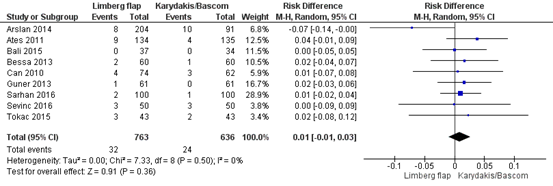
Ten studies reported on the outcome recurrence. Eight studies reported the recurrence rate with a follow-up period of at least 12 months, of which six compared the Limberg flap with the Karydakis flap (Arslan, 2014; Ates, 2011; Bali, 2015; Bessa, 2013; Can, 2010; Sevinç, 2016), and two with Bascom cleft lift (Guner, 2013; Sarhan, 2016). In total, 1399 patients were analysed, with 763 in the Limberg flap group, and 636 in the Karydakis/Bascom procedure group. In the Limberg flap group, 32/763 (4.2%) had a recurrence, compared to 24/636 (3.8%) in the Karydakis/Bascom group. The pooled risk difference is 0.01 (95% CI: -0.01 to 0.03), favouring the Karydakis/Bascom procedure, which is not clinically relevant (Figure 1).
Two studies reported the recurrence rate with 6 months follow-up (Alvandipour, 2019; Riaz, 2019). Alvandipour (2019) reported 0/27 (0%) recurrences in the Limberg flap group and 1/37 (3%) in the Karydakis flap group (risk difference: -0.03, 95% CI: -0.11 to 0.05), favouring Limberg flap procedure. Riaz (2019) reported 9/102 (9%) recurrences in the Limberg flap group, and 5/102 (5%) in the Karydakis procedure group (risk difference: 0.04, 95% CI: -0.03 to 0.11), favouring the Karydakis procedure. These studies were not taken into account in the pooled analyses, because of the short follow-up period.
Figure 1 Outcome recurrence rate (>12 months follow-up) with Limberg flap versus Karydakis/Bascom procedure
Z: p-value of pooled effect; df: degrees of freedom, I2: statistical heterogeneity, CI: confidence interval
Return to daily activities (important)
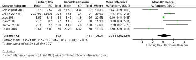
Eight studies reported on the outcome return to daily activities, of which six reported the mean and standard deviation (Alvandipour, 2019; Arslan, 2014; Ates, 2011; Can, 2010; Sarhan, 2016; Tokac, 2015). In total, 1074 patients were analysed, with 593 patients in the Limberg flap group, and 481 in the Karydakis/Bascom procedure group. The pooled mean difference in days until return to daily activities was 0.24 days (95% CI: -1.05 to 1.52), favouring the Karydakis/Bascom procedure, which is not clinically relevant (figure 2).
Two studies reported the median time to return to daily activities and were therefore not taken into account in the pooled analyses, but only described here. Bali (2015) reported a median of 8 days (6 – 12, not specified whether this was a range or interquartile range) in the Limberg flap group and 17 (14 – 20) in the Karydakis procedure group, and Ersoy (2009) reported a median of 14 days (range: 5 – 46) in the Limberg flap and 15 days (range: 5 – 45) until return to daily activities in the Karydakis procedure group.
Figure 2 Outcome return to daily activities with Limberg flap versus Karydakis/Bascom procedure
Z: p-value of pooled effect; df: degrees of freedom, I2: statistical heterogeneity, CI: confidence interval
Quality of life (important)
One study reported the outcome quality of life, measured with the SF-36, on day ten and day 30 (Guner, 2013), see table 1. They reported: “the SF-36 survey showed statistically significant differences between the two groups on postoperative day ten in regard to bodily pain and role limitations due to physical problems. The results of these variables were higher in the Bascom cleft lift patients. A statistically significant difference was not observed between the other parameters.
Table 1 – Quality of life scores, measured with the SF-36
|
Subscale / postop day |
Limberg flap group |
Bascom cleft lift group |
p-value |
|
Physical functioning |
|
|
|
|
Day 10 |
79.59 ± 18.17 |
80.41 ± 17.09 |
0.93 |
|
Day 30 |
92.29 ± 10.31 |
94.67 ± 8.74 |
0.18 |
|
Role-physical |
|
|
|
|
Day 10 |
41.80 ± 30.86 |
54.92 ± 34.10 |
0.03 |
|
Day 30 |
76.64 ± 35.02 |
83.61 ± 26.17 |
0.41 |
|
Bodily pain |
|
|
|
|
Day 10 |
66.77 ± 18.41 |
76.23 ± 20.53 |
0.02 |
|
Day 30 |
83.87 ± 14.48 |
84.06 ± 21.66 |
0.18 |
|
General health |
|
|
|
|
Day 10 |
76.73 ± 26.65 |
83.2 ± 15.65 |
0.49 |
|
Day 30 |
84.87 ± 22.29 |
89.79 ± 10.79 |
0.88 |
|
Vitality |
|
|
|
|
Day 10 |
75.98 ± 15.57 |
70.16 ± 17.77 |
0.10 |
|
Day 30 |
78.74 ± 13.61 |
74.02 ± 17.95 |
0.18 |
|
Social functioning |
|
|
|
|
Day 10 |
71.11 ± 23.33 |
71.72 ± 22.11 |
0.93 |
|
Day 30 |
80.12 ± 18.87 |
83.4 ± 13.64 |
0.54 |
|
Role-emotional |
|
|
|
|
Day 10 |
57.38 ± 30.52 |
56.83 ± 36.69 |
0.95 |
|
Day 30 |
78.69 ± 24.37 |
83.07 ± 20.74 |
0.37 |
|
Mental health |
|
|
|
|
Day 10 |
79.67 ± 13.69 |
79.11 ± 13.75 |
0.59 |
|
Day 30 |
79.15 ± 15.36 |
77.11 ± 13.95 |
0.14 |
Data are expressed as the mean ± SD. Higher scores indicate better health status. Surgical complications (important)
Wound infection
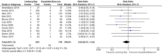
Eleven studies reported on the outcome wound infection (Alvandipour, 2019; Arslan, 2014; Ates, 2011; Bali, 2015; Bessa, 2013; Can, 2010; Ersoy, 2009; Guner, 2013; Riaz, 2019; Sevinç, 2016; Tokac, 2015). In total, 1575 patients were analysed, with 845 in the Limberg flap group and 730 in the Karydakis/Bascom procedure group. In the Limberg flap group, 51/845 (6.0%) had a wound infection, and in the Karydakis/Bascom procedure group, 55/730 (7.5%) had a wound infection. The pooled risk ratio is 0.84 (95% CI: 0.57 to 1.24), favouring the Limberg flap, which is not clinically relevant (figure 3).
Figure 3 Outcome wound infection with Limberg flap versus Karydakis/Bascom procedure
Z: p-value of pooled effect; df: degrees of freedom, I2: statistical heterogeneity, CI: confidence interval
Wound dehiscence
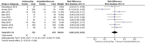
Nine studies reported on the outcome wound dehiscence infection (Alvandipour, 2019; Arslan, 2014; Ates, 2011; Bali, 2015; Can, 2010; Guner, 2013; Riaz, 2019; Sarhan, 2016; Sevinç, 2016). In total, 1470 patients were analysed, with 792 in the Limberg flap group and 678 in the Karydakis/Bascom procedure group. In the Limberg flap group, 47/792 (5.9%) had wound dehiscence, and in the Karydakis/Bascom procedure group 40/678 (5.9%) had wound dehiscence. The pooled risk difference is 0.00 (95% CI: -0.03 to 0.03), favouring neither intervention, which is not clinically relevant (figure 4).
Figure 4 Outcome wound dehiscence with Limberg flap versus Karydakis/Bascom procedure
Z: p-value of pooled effect; df: degrees of freedom, I2: statistical heterogeneity, CI: confidence interval
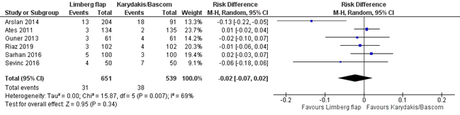
Seroma
Six studies reported on the outcome seroma (Arslan, 2014; Ates, 2011; Guner, 2013; Riaz, 2019; Sarhan, 2016; Sevinç, 2016). In total, 1190 patients were analysed, with 651 in the Limberg flap group and 539 in the Karydakis/Bascom procedure group. All patient were treated with a suction drain postoperatively. In the Limberg flap group, 31/651 (5%) had seroma, and in the Karydakis/Bascom procedure group 38/539 (7%) had seroma. The pooled risk difference is -0.02 (95% CI: -0.07 to 0.02), favouring Limberg flap, which is not clinically relevant (figure 5).
Figure 5 Outcome seroma with Limberg flap versus Karydakis/Bascom procedure
Z: p-value of pooled effect; df: degrees of freedom, I2: statistical heterogeneity, CI: confidence interval
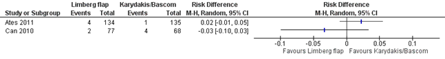
Complications that required re-operation
Two studies reported on the outcome complications that required a re-operation (Ates, 2011; Can, 2010). In total, 414 patients were analysed, with 211 in the Limberg flap and 203 in the Karydakis/Bascom procedure group. In the Limberg flap group, 6/211 (3%) had complications that required a re-operation, and in the Karydakis/Bascom procedure group 5/203 (2%) had complications that required a re-operation. Complications that required reoperation were wound dehiscence or wound infection that needed removal of the stitches and secondary wound healing. Because there were only two studies that reported this outcome, the results were not pooled, and the clinical relevance could not be determined.
Figure 6 Outcome complications that required re-operation with Limberg flap versus Karydakis/Bascom procedure
Z: p-value of pooled effect; df: degrees of freedom, I2: statistical heterogeneity, CI: confidence interval
Postoperative pain (important)
Six studies reported on the outcome postoperative pain (Alvandipour, 2019; Ates, 2011; Bali, 2015; Ersoy, 2009; Guner, 2013; Sarhan, 2016). Due to the different scales used for the Visual Analogous Scale (VAS), timepoints at which pain was assessed, and different ways of data presentation (mean and standard deviation, or median and range) it was not possible to pool the data. Therefore, the data is presented in table 2.
Table 2 – Pain scores measured with VAS
|
|
Limberg flap |
Karydakis/Bascom procedure |
|
Alvandipour (2019) |
Pain while sitting on the operation site (1-10), mean ± sd |
|
|
Timing unknown |
4.00 ± 0.96 |
4.11 ± 1.02 |
|
Ates (2011) |
Pain of operation site (1-10), mean ± sd |
|
|
Day 15 |
5.56 ± 1.55 |
5.58 ± 1.52 |
|
Day 30 |
3.23 ± 1.14 |
2.22 ± 1.01 |
|
Bali (2015) |
Pain (scale not reported), unit not reported |
|
|
Timing unknown |
2 (1–3) |
4 (2–6) |
|
Ersoy (2009) |
Pain (1-10), median (min-max) |
|
|
At discharge |
4 (0–9) |
4 (0–10) |
|
Guner (2013) |
Pain (0-100), mean ± sd |
|
|
Day 2 |
9.31 ± 10.53 |
8.39 ± 15.05 |
|
Day 10 |
3.97 ± 5.64 |
2.62 ± 4.79 |
|
Day 20 |
1.21 ± 2.44 |
0.95 ± 2.36 |
|
Sarhan (2016) |
Pain (0-10), mean ± sd |
|
|
During follow-up |
3.4 ± 1.5 |
3.7 ± 1.6 |
Level of evidence of the literature
Recurrence (crucial)
The certainty of the evidence started high, as the evidence originated from RCTs. The level of evidence regarding the outcome measure recurrence was downgraded by one level for risk of bias (mostly no intention-to-treat analyses and blinding), and one level for imprecision (low number of events and confidence interval includes ‘no effect’). The level of evidence was therefore graded as low.
Return to daily activities (important)
The certainty of the evidence started high, as the evidence originated from RCTs. The level of evidence regarding the outcome return to daily activities was downgraded by one level for risk of bias (mostly no intention-to-treat analyses and blinding), one level for inconsistency (conflicting results across studies), one level for imprecision (low number of patients and confidence interval includes ‘no effect’). The level of evidence was therefore graded as very low.
Quality of life (important)
The certainty of the evidence started high, as the evidence originated from RCTs. The level of evidence regarding the outcome quality of life was downgraded by one level for risk of bias (mostly no intention-to-treat analyses and blinding), two levels for imprecision (very low number of patients). The level of evidence was therefore graded as very low.
Surgical complications (important)
Wound infection
The certainty of the evidence started high, as the evidence originated from RCTs. The level of evidence regarding the outcome wound infection was downgraded by one level for risk of bias (mostly no intention-to-treat analyses and blinding), one level for inconsistency (inconsistent results) and one level for imprecision (low number of patients). The level of evidence was therefore graded as low.
Wound dehiscence
The certainty of the evidence started high, as the evidence originated from RCTs. The level of evidence regarding the outcome wound dehiscence was downgraded by one level for risk of bias (mostly no intention-to-treat analyses and blinding), one level for inconsistency (inconsistent results) and one level for imprecision (low number of patients). The level of evidence was therefore graded as low.
Seroma
The certainty of the evidence started high, as the evidence originated from RCTs. The level of evidence regarding the outcome seroma was downgraded by one level for risk of bias (mostly no intention-to-treat analyses and blinding), one level for inconsistency (conflicting results) and one level for imprecision (low number of patients). The level of evidence was therefore graded as low.
Complications that required re-operation
The certainty of the evidence started high, as the evidence originated from RCTs. The level of evidence regarding the outcome complications that required re-operation was downgraded by one level for risk of bias (no intention-to-treat analyses and blinding), one level for inconsistency (conflicting results) and two levels for imprecision (very low number of patients). The level of evidence was therefore graded as very low.
Postoperative pain (important)
The certainty of the evidence started high, as the evidence originated from RCTs. The level of evidence regarding the outcome postoperative pain was downgraded by one level for risk of bias (mostly no intention-to-treat analyses and blinding), two levels for imprecision (very low number of patients). The level of evidence was therefore graded as very low.
A systematic review of the literature was performed to answer the following question:
P: Patients with symptomatic pilonidal sinus
I: Excision with closure by flap repair (primary healing), with techniques that cross the midline ((modified) Limberg flap, Dufourmentel, V-Y plasty, Z-plasty, Rhomboid flap, advancement flap, rotation flap, transposition flap)
C: Excision with closure by flap repair (primary healing), with off-midline closure (Karydakis, Bascom cleft lift)
O: Recurrence, return to daily activities, quality of life, pain, surgical complications (wound infection, wound dehiscence, seroma, complication that required re-operation)
Relevant outcome measures
The guideline development group considered recurrence as a crucial outcome measure for decision making; and surgical complications, postoperative pain, return to daily activities, and quality of life as an important outcome measure for decision making.
The working group defined the outcome measures as follows:
- Recurrence: a healed surgical site with de novo midline pits/sinus/secondary sinus opening after a symptom free period. Total recurrence rate of the study should be measured preferably with a study follow-up of at least 12 months.
- Return to daily activities: time from surgery to first day of return to daily activity (including work, sports, study or other activities).
- Quality of life: quality of life measured with a validated questionnaire.
- Surgical complications: wound infection, wound dehiscence, seroma or any complication that required re-operation.
- Postoperative pain: postoperative pain measured with a Visual Analog Scale (VAS) or the reported use of analgetics.
The guideline development group used the GRADE standard limits of 25% for dichotomous outcome measures as a minimal clinically (patient) important difference. Risk ratios were described where possible. In case studies that reported no events in one of the groups, risk differences were used. A risk difference of 10% was considered clinically relevant.
The following limits were used for minimal clinically (patient) important differences per outcome:
- Recurrence: GRADE standard limits of 25% for dichotomous outcome measures (RR <0.80 or RR >1.25)
- Return to daily activities: 1 working week (5 days)
- Quality of life: 0.5 sd
- Surgical complications: GRADE standard limits of 25% for dichotomous outcome measures (RR <0.80 or RR >1.25)
- Postoperative pain: 2 points on a VAS-scale.
Search and select (Methods)
The databases Medline (via OVID) and Embase (via Embase.com) were searched with relevant search terms until April 22nd, 2021. The detailed search strategy is depicted under the tab Methods. The systematic literature search resulted in 52 hits. Studies were selected based on the following criteria: included patients with symptomatic pilonidal disease, compared midline flap repair techniques with off-midline flap repair techniques, reported at least one of the outcomes of interest, the study design is a systematic review (SR) of randomised controlled trials (RCTs) or RCT, and was written in English language.
Thirty-nine studies (14 SRs and 25 RCTs) were initially selected based on title and abstract screening. After reading the full text, 35 studies were excluded (see the table with reasons for exclusion under the tab Methods), and four studies (1 SR and 3 RCTs) were included.
- Alvandipour, M., Zamani, M. S., Ghorbani, M., Charati, J. Y., & Karami, M. Y. (2019). Comparison of Limberg flap and Karydakis flap surgery for the treatment of patients with pilonidal sinus disease: a single-blinded parallel randomized study. Annals of coloproctology, 35(6), 313.
- Arslan, K., Kokcam, S. S., Koksal, H., Turan, E., Atay, A., & Dogru, O. (2014). Which flap method should be preferred for the treatment of pilonidal sinus? A prospective randomized study. Techniques in coloproctology, 18(1), 29-37.
- Ates, M., Dirican, A., Sarac, M., Aslan, A., & Colak, C. (2011). Short and long-term results of the Karydakis flap versus the Limberg flap for treating pilonidal sinus disease: a prospective randomized study. The American Journal of Surgery, 202(5), 568-573.
- Bali, İ., Aziret, M., Sözen, S., Emir, S., Erdem, H., Çetinkünar, S., & İrkörücü, O. (2015). Effectiveness of Limberg and Karydakis flap in recurrent pilonidal sinus disease. Clinics, 70, 350-355.
- Bessa, S. S. (2013). Comparison of short-term results between the modified Karydakis flap and the modified Limberg flap in the management of pilonidal sinus disease: a randomized controlled study. Diseases of the colon & rectum, 56(4), 491-498.
- Can, M. F., Sevinc, M. M., Hancerliogullari, O., Yilmaz, M., & Yagci, G. (2010). Multicenter prospective randomized trial comparing modified Limberg flap transposition and Karydakis flap reconstruction in patients with sacrococcygeal pilonidal disease. The American journal of surgery, 200(3), 318-327.
- Guner, A., Boz, A., Ozkan, O. F., Ileli, O., Kece, C., & Reis, E. (2013). Limberg flap versus Bascom cleft lift techniques for sacrococcygeal pilonidal sinus: prospective, randomized trial. World journal of surgery, 37(9), 2074-2080.
- Ray, K., Albendary, M., Baig, M. K., Swaminathan, C., Sains, P., & Sajid, M. S. (2020). Limberg flap for the management of pilonidal sinus reduces disease recurrence compared to Karydakis and Bascom procedure: a systematic review and meta-analysis of randomized controlled trials. Minerva Chirurgica, 75(5), 355-64.
- Riaz, A., Afzal, A., & Dogar, M. A. (2019). Comparison of Limberg Flap with Karydakis Repair in Pilonidal Sinus Disease. Med Forum 30(10), 91.
- Sarhan, A. E., Sherif, T., & Zakaria, Y. (2016). A prospective randomized trial comparing modified Limberg flap and cleft lift procedure in the treatment of uncomplicated sacrococcygeal pilonidal disease. The Egyptian Journal of Surgery, 35(2), 89.
- Sevinç, B., Karahan, Ö., Okuş, A., Ay, S., Aksoy, N., & Şimşek, G. (2016). Randomized prospective comparison of midline and off-midline closure techniques in pilonidal sinus surgery. Surgery, 159(3), 749-754.
- Stauffer, V. K., Luedi, M. M., Kauf, P., Schmid, M., Diekmann, M., Wieferich, K., ... & Doll, D. (2018). Common surgical procedures in pilonidal sinus disease: a meta-analysis, merged data analysis, and comprehensive study on recurrence. Scientific reports, 8(1), 1-28.
- Tokac, M., Dumlu, E. G., Aydin, M. S., Yalcın, A., & Kilic, M. (2015). Comparison of modified Limberg flap and Karydakis flap operations in pilonidal sinus surgery: prospective randomized study. International surgery, 100(5), 870-877.
Evidence table for systematic review of RCTs and observational studies (intervention studies)
Research question: Welk type verschuivingsplastiek heeft de voorkeur als men besluit tot excisie met verschuivingsplastiek?
|
Study reference |
Study characteristics |
Patient characteristics |
Intervention (I) |
Comparison / control I
|
Follow-up |
Outcome measures and effect size |
Comments |
|
Ray, 2020
PS., study characteristics and results are extracted from the SR (unless stated otherwise) |
SR and meta-analysis of RCTs
Literature search up to November 2019
A: Arslan, 2014 B: Ates, 2011 C: Bali, 2015 D: Bessa, 2013 E: Can, 2010 F: Ersoy, 2009 G: Guner, 2013 H: Sarhan, 2016 I: Sevinc, 2016 J: Tokac, 2015
Study design: A to J: RCT
Setting and Country: A: Turkey B: Turkey C: Turkey D: Egypt E: Turkey F: Turkey G: Turkey H: Egypt I: Turkey J: Turkey
Source of funding and conflicts of interest: Source of funding not reported. The authors certify that there is no conflict of interest with any financial organization regarding the material discussed in the manuscript. |
Inclusion criteria SR: (1) randomised controlled trial (2) comparing LF versus excision of sinus pilonidalis± closure/Karydakis procedure/ Bascom procedure.
Exclusion criteria SR: No exclusion criteria were applied.
18 studies included, of which 10 met our PICO
Important patient characteristics at baseline:
N, intervention / control group A: 204 / 91 (I: LF: 96, MLF: 108) B: 134 / 135 C: 37 /34 D: 60 / 60 E: 77 / 68 F: 50 / 50 G: 61 / 61 H: 100 / 100 I: 50 / 50 J: 46 / 46
A: LF: 26.5 ± 5.9, MLF: 24.7 ± 5.1, C: 24.7 ± 5.1 B: I: 25.50 (7.11), C: 24.45 (7.30) C: I: 25, C: 23.5 D: I: 23 (16–41), C: 23 (15–36) E: I: 22 (19–40), C: 22 (20–31) F: I: 25.8 ± 6.4, C: 27.8 ± 6.85 G: I: 25.41 (18–48), C: 24.64 (18–46) H: I: 23 (18-44), C: 22 (19-52) I: I: 23.5 (18–46), C: 24 (18–38) J: I: 29.28 6 8.53, C: 28.35 6 8.48
Sex: (n/N (%) male) A: LF: 68/96 (71%), MLF: 95/108 (88%), C: 77/91 (85%) B: 117/134 (87%), C: 123/135 (91%) C: I: 31/37 (86%), C: 32/34 (94%) D: I: 58/60 (97%), C: 54/60 (90%) E: I: 76/77 (99%), C: 67/68 (99%) F: 32/50 (64%), C: 36/50 (72%) G: I: 48/61 (79%), C: 50/61 (82%) H: I: 80/100 (80%), C: 75/100 (75%) I: I: 39/50 (78%) C: 41/50 (82%), J: I: 40/46 (87%), C: 39/46 (87%)
Groups comparable at baseline? Not reported. |
A: 1) Limberg flap, 2) modified Limberg flap B: Limberg flap C: Limberg flap D: modified Limberg flap E: modified Limberg flap F Limberg flap G: Limberg flap H: modified Limberg flap I: Limberg flap J: modified Limberg flap
* extracted from individual studies
|
A: Karydakis flap B: Karydakis flap C: Karydakis flap D: modified Karydakis flap E: Karydakis flap F: Karydakis flap G: Bascom cleft lift H: Bascom cleft lift I: Karydakis flap J: Karydakis flap
* extracted from individual studies |
End-point of follow-up:* A: 33 (24-42) B: 26.40 ± 8.2 C: 28 D: 20.5 E: 16.8 ± 3.4 F: 1 G: 13 H: I: 21.5 ± 6.8 / C: 22 ± 7.6 I: 24.2 (18.5-34.27) J: 26 (16-28)
* extracted from individual studies
For how many participants were no complete outcome data available? Not reported.
|
Effect measure: RD [95% CI]: A: I: 8/204, C: 10/91 B: I: 9/134, C: 4/135 C: I: 0/37, C: 0/34 D: I: 2/60, C: 1/60 E: I: 4/74, C: 3/63 F: not reported G: I: 1/61, C: 0/61 H: I: 2/100, C: 1/100 I: I: 3/50, C: 3/50 J: I: 3/43, C: 2/43
Pooled effect (random effects model): Risk difference 0.01 [95% CI -0.01 to 0.03] favouring Karydakis flap Heterogeneity (I2): 0%
Return to daily activities * A: I(1): 20.8 ± 6.5, I(2): 19.8 ± 4.6, C: 19.1 ± 3.4 B: I: 8.55 ± 1.18, C: 8.45 ± 1.16 C: I: 8 (6–12), C: 17 (14–20) D: not reported E: I: 21.5 ± 8.5, C: 18.8 ± 7.7 F: I: 14 (5–46), C: 15 (5–45) G: not reported H: I: 21.4 ± 7.5, C: 18.7 ± 7.6 I: not reported J: I: 20.61 ± 7.89, C: 23.29 ± 6.42
Pooled effect (random effects model): mean difference: 0.87 days [95% CI -0.25 to 1.99] favouring Karydakis/Bascom Heterogeneity (I2): 71%
Quality of life * A: not reported B: not reported C: not reported D: not reported E: not reported F: not reported G: Day 10: I: 79.59 ± 18.17, C: 80.41 ± 17.09; Day 30: I: 92.29 ± 10.31, C: 94.67 ± 8.74 Day 10: I: 76.73 ± 26.65, C: 83.2 ± 15.65; Day 30: I: 84.87 ± 22.29, C: 89.79 ± 10.79. Other subscales: see Table 2 of the paper. H: not reported I: not reported J: not reported
Surgical complications * Wound infection A: I: 7/204, C: 6/91 B: I: 8/134, C: 4/135 C: I: 4/37, C: 8/34 D: I 3/60, C: 2/60 E: I: 1/77, C: 0/68 (during in-hospital period), I: 3/77, C: 3/68 (during outpatient visit on day 30) F: I: 4/50, C: 13/50 G: I: 5/61, C: 5/61 (superficial); 1/61, C: 0/61 (deep) H: not reported I: I: 2/50, C: 1/50 J: I: 3/43, C: 3/42
Pooled effect (random effects model): RR: 0.80 [95% CI 0.51 to 1.27] favouring Karydakis/Bascom Heterogeneity (I2): 11%
Wound dehiscence A: 6/204, C: 14/91 (partial wound dehiscence) B: I: 14/134, C: 8/135 C: I: 1/37, C: 2/34 D: not reported E: I: 1/77, C: 1/68 (during in-hospital period); I: 2/77, C: 2/68 (during outpatient visit postop day 30) F: not reported G: I: 3/61, C: 2/61 (partial); I: 0/61, C: 1//61 (total) H: I: 4/100, C: 4/100 I: I: 1/50, C: 0/50 J: not reported
Pooled effect (random effects model): risk difference: -0.01 [95% CI -0.04 to 0.03] favouring Limberg flap Heterogeneity (I2): 55%
Seroma A: I: 13/204, C: 18/91 B: I: 3/134, C: 2/135 C: not reported D: not reported E: not reported F not reported G: I: 3/61, C: 4/61 H: I: 5/100, C: 3/100 (seroma and/or hematoma) I: I: 4/50, C: 7/50 J: not reported
Pooled effect (random effects model): risk difference: -0.03 [95% CI -0.09 to 0.03] favouring Limberg flap Heterogeneity (I2): 76%
Complications that required re-operation A: not reported B: I: 4/134, C: 1/135 C: not reported D: not reported E: I: 2/77, C: 4/68 F: not reported G: not reported H: not reported I: not reported J: not reported
Pooled effect (random effects model): xx xxx [95% CI xx to xx] favouring xx Heterogeneity (I2): xx%
Postoperative pain * A: not reported B: VAS score for pain of operation site: Day 15: I: 5.56 ± 1.55, C: 5.58 ± 1.52. Day 30: I: 3.23 ± 1.14, C: 2.22 ± 1.01 C: VAS score: I: 2 (1–3), C: 4 (2–6) D: not reported E: not reported F: VAS score: I: 4 (0–9), C: 4 (0–10) G: VAS score: day 2: I: 9.31 ± 10.53, C: 8.39 ± 15.05; day 10: I: 3.97 ± 5.64, C: 2.62 ± 4.79; day 20: I: 1.21 ± 2.44, C: 0.95 ± 2.36 H: VAS score: I: 3.4 ± 1.5, C: 3.7 ± 1.6 I: not reported J: not reported
* extracted from individual studies |
Remarks - The SR included studies with LF versus excision of PNS ± closure/Karydakis procedure/Bascom procedure. We only included studies comparing LF with Karydakis or Bascom II procedures. - 8/10 studies were performed in Turkey, and 2/10 in Egypt
Brief description of author’s conclusion Limberg flap seems to have clinical advantage over Karydakis and Bascom procedure in terms of reduced recurrence rate following surgical excision of pilonidal sinus. Although, this advantage was clinically persisted on subgroup analysis but failed to achieve statistical significance.
|
Evidence table for intervention studies
Research question: Welk type verschuivingsplastiek heeft de voorkeur als men besluit tot excisie met verschuivingsplastiek?
|
Study reference |
Study characteristics |
Patient characteristics 2 |
Intervention (I) |
Comparison / contrI(C) 3
|
Follow-up |
Outcome measures and effect size 4 |
Comments |
|
Alvandipour, 2019
|
Type of study: RCT, single blind.
Setting and country: Imam Khomeini Hospital (Sari), Iran.
Funding and conflicts of interest: Funding was not reported. Conflict of interest: none reported.
|
Inclusion criteria: (1) patients with intergluteal pilonidal disease who had been referred to Imam Khomeini Hospital (2) whose ages ranged from 15 to 65 years.
Exclusion criteria: (1) elderly patients with comorbid diseases (2) scars from a previous pilonidal surgery (3) abscess presentations (4) diabetes mellitus (5) immunodeficiency (6) neurological disorders (7) drug addiction or alcoholism (8) American Society of Anesthesiologists physical status classification grade III–IV, (9) age < 15 years or > 65 years (10) orifice located more than 3 cm from the sinus center.
N total at baseline: Intervention: 28 Control: 37
Important prognostic factors2: Age (mean ± SD): I: 34.2 ± 10.5 C: 25.9 ± 9.5
Sex (n/N (%) men) I: 18/27 (67%) C: 16/37 (43%)
Groups comparable at baseline? Patients in the control group were statistically significant younger than intervention group. Other baseline characteristics were not reported.
|
Limberg flap
Surgery was performed in the LF group, as defined by Mentes et al. A rhomboid excision was performed with the lower edge 2 cm lateral to the midline and covered the entire area where the sinus extended. Hemostasis was accomplished by using electrocautery. In order to ensure tension-free repair, the flap was released at the bottom involving the gluteal fascia and was then glided medially to cover the cavity defect. A suction drain was applied to the region in all patients. The subcutaneous tissue was closed with 2 fold 2/0 polyglactin suture (Ethicon US, LLC, Cincinnati, OH, USA), and the skin was closed with a 3/0 polypropylene mattress suture (Ethicon US, LLC). The drain was removed when drainage fell below 40 mL/day.
|
Karydakis flap
Surgery was performed in the KF group, as described by Karydakis. In this technique, an asymmetrical elliptic excision was made with the lower and upper edges located approximately 2 cm lateral to the natal cleft; all defective tissues were removed until the healthy borders had been reached. Afterwards, the medical wound edge was mobilized, and the flap was slid by suturing it to the fascia. The subcutaneous tissue was closed with 2 fold 2/0 polyglactin suture, and the skin was closed with a 3/0 polypropylene mattress suture. |
Length of follow-up: 6 months
Loss-to-follow-up: Intervention: 1/28 (4%) Reasons not provided.
Control: 0/37 (0%) Reasons not applicable.
Incomplete outcome data: Intervention: 1/28 (4%) Reasons: lost to follow-up.
Control: 0/37 (0%) Reasons not applicable.
|
Not reported with >12 months follow-up.
Recurrence with 6 months follow-up I: 0/27 (0%) C: 1/37 (3%)
Return to daily activities I: 9.15 ± 2.52 C: 11.59 ± 3.44
Not reported.
Surgical complications Wound infection I: 1/27 (4%) C:3/37 (8%)
Wound dehiscence I: 0/27 (0%) C: 1/37 (3%)
Seroma Not reported.
Aany complication that requires re-operation Not reported.
VAS pain score (mean ± SD) I: 4.00 ± 0.96 C: 4.11 ± 1.02
|
No intention-to-treat analyses were performed.
Author’s conclusion: Limberg flap surgery is the standard choice for the treatment of patients with pilonidal disease. Compared to Karydakis flap surgery, it has fewer complications, faster return to work, better overall patient satisfaction, and shorter complete wound healing time. |
|
Riaz, 2019
|
Type of study: RCT
Setting and country: Central Park Teaching Hospital Lahore, Pakistan.
Funding and conflicts of interest: Funding not reported. Conflict of interest: none reported. |
Inclusion criteria: (1) patients of either gender presenting with saccrococcygeal pilonidal sinus (2) age ranging from 15 to 60 years (3) ASA grade I to III
Exclusion criteria: (1) patients who presented with a concurrent abscess (2) recurrent pilonidal sinus (3) concurrent perianal pathology like fistula in ano (4) ASA grade IV and V (5) diabetes mellitus (6) compromised immune status
N total at baseline: Intervention: 102 Control: 102
Important prognostic factors2: Age (mean ± SD): I: 28.1 ± 8.7 C: 26.4 ± 7.7
Sex (n/N (%) men) I: 86/102 (84%) C 80/102 (78%)
Groups comparable at baseline? Not reported. |
Limberg flap technique
A rhombus shaped area including the pilonidal sinus tract was excised up till the pre-sacral fascia. A rhombus shaped tension free fasciocutaneous flap was then placed to cover the defect.
|
Karydakis repair technique
An elliptical D shaped incision was given and extended down till pre-sacral fascia to excise the pilonidal sinus tract. A double layered closure was then done with the suture line lying away from midline |
Length of follow-up: 6 months
Loss-to-follow-up: not reported
Incomplete outcome data: Not reported.
|
Not reported with >12 months follow-up.
Recurrence with 6 months follow-up I: 9/102 (9%) C: 5/102 (5%)
Return to daily activities Not reported.
Not reported
Surgical complications Wound infection I: 9/102 (9%) C:7/102 (7%)
Wound dehiscence I: 15/102 (15%) C: 6/102 (6%)
Seroma I: 3/102 (3%) C: 4/102 (4%)
Aany complication that requires re-operation Not reported
Pain VAS score on the 28th postoperative day I: 2.51 ± 1.16 C: 3.19 ± 1.45
|
Remarks - Not reported whether there were patients lost to follow-up and analyses were intention-to-treat. - Method of randomisation not clearly described.
Author’s conclusion: Both Limberg flap and Karydakis repair are effective surgical options for the management of pilonidal sinus disease. The two techniques have a comparable complication and recurrence rate. However Karydakis repair stands out as the better technique in terms of lesser operation time and lesser frequency of wound dehiscence while Limberg flap technique is associated with lesser post-operative pain. |
Notes:
- Prognostic balance between treatment groups is usually guaranteed in randomized studies, but non-randomized (observational) studies require matching of patients between treatment groups (case-control studies) or multivariate adjustment for prognostic factors (confounders) (cohort studies); the evidence table should contain sufficient details on these procedures
- Provide data per treatment group on the most important prognostic factors [(potential) confounders]
- For case-control studies, provide sufficient detail on the procedure used to match cases and controls
- For cohort studies, provide sufficient detail on the (multivariate) analyses used to adjust for (potential) confounders
Risk of bias assessment
Table of quality assessment for systematic reviews of RCTs and observational studies
Research question: Welk type verschuivingsplastiek heeft de voorkeur als men besluit tot excisie met verschuivingsplastiek?
|
Study
First author, year |
Appropriate and clearly focused question?1
Yes/no/unclear |
Comprehensive and systematic literature search?2
Yes/no/unclear |
Description of included and excluded studies?3
Yes/no/unclear |
Description of relevant characteristics of included studies?4
Yes/no/unclear |
Appropriate adjustment for potential confounders in observational studies?5
Yes/no/unclear/not applicable |
Assessment of scientific quality of included studies?6
Yes/no/unclear |
Enough similarities between studies to make combining them reasonable?7
Yes/no/unclear |
Potential risk of publication bias taken into account?8
Yes/no/unclear |
Potential conflicts of interest reported?9
Yes/no/unclear |
|
Ray, 2020 |
Yes |
Yes |
Yes |
Yes |
Not applicable |
Yes |
Yes |
No |
Yes |
- Research question (PICO) and inclusion criteria should be appropriate and predefined
- Search period and strategy should be described; at least Medline searched; for pharmacological questions at least Medline + EMBASE searched
- Potentially relevant studies that are excluded at final selection (after reading the full text) should be referenced with reasons
- Characteristics of individual studies relevant to research question (PICO), including potential confounders, should be reported
- Results should be adequately controlled for potential confounders by multivariate analysis (not applicable for RCTs)
- Quality of individual studies should be assessed using a quality scoring tool or checklist (Jadad score, Newcastle-Ottawa scale, risk of bias table etc.)
- Clinical and statistical heterogeneity should be assessed; clinical: enough similarities in patient characteristics, intervention and definition of outcome measure to allow pooling? For pooled data: assessment of statistical heterogeneity using appropriate statistical tests (e.g. Chi-square, I2)?
- An assessment of publication bias should include a combination of graphical aids (e.g., funnel plot, other available tests) and/or statistical tests (e.g., Egger regression test, Hedges-Olken). Note: If no test values or funnel plot included, score “no”. Score “yes” if mentions that publication bias could not be assessed because there were fewer than 10 included studies.
- Sources of support (including commercial co-authorship) should be reported in both the systematic review and the included studies. Note: To get a “yes,” source of funding or support must be indicated for the systematic review AND for each of the included studies.
Risk of bias table for intervention studies (randomized controlled trials)
Research question: Welk type verschuivingsplastiek heeft de voorkeur als men besluit tot excisie met verschuivingsplastiek?
|
Study reference
(first author, publication year) |
Describe method of randomisation1 |
Bias due to inadequate concealment of allocation?2
(unlikely/likely/unclear) |
Bias due to inadequate blinding of participants to treatment allocation?3
(unlikely/likely/unclear) |
Bias due to inadequate blinding of care providers to treatment allocation?3
(unlikely/likely/unclear) |
Bias due to inadequate blinding of outcome assessors to treatment allocation?3
(unlikely/likely/unclear) |
Bias due to selective outcome reporting on basis of the results?4
(unlikely/likely/unclear) |
Bias due to loss to follow-up?5
(unlikely/likely/unclear) |
Bias due to violation of intention to treat analysis?6
(unlikely/likely/unclear) |
|
Alvandipour, 2019 |
A computer-based table of randomization was used. |
Note: sealed envelopes were used. |
Note: Single-blind study; patients, outcome assessor and data analyst were blinded, main researcher (surgeon) was not blinded. |
Unlikely
Note: Single-blind study; patients, outcome assessor and data analyst were blinded, main researcher (surgeon) was not blinded. |
Unlikely
Note: Single-blind study; patients, outcome assessor and data analyst were blinded, main researcher (surgeon) was not blinded. |
Note: alle relevant outcomes were reported |
Unlikely
Note: minimal loss to follow-up, reasons were given. |
Note: No ITT analyses were performed. |
|
Riaz, 2019 |
Patients were randomly divided into two equal groups of n patients each by lottery method” |
Unclear
Note: method of allocation concealment was not described. |
It was not reported whether patients, healthcare provides, data collectors, outcome assessor and data analysts were blinded. |
Unclear
It was not reported whether patients, healthcare provides, data collectors, outcome assessor and data analysts were blinded. |
Unclear
It was not reported whether patients, healthcare provides, data collectors, outcome assessor and data analysts were blinded. |
Unlikely
Note: alle relevant outcomes were reported |
Unclear
Note: not reported whether there was loss to follow-up. |
Unclear
Note: not reported whether ITT analyses were performed. |
- Randomisation: generation of allocation sequences have to be unpredictable, for example computer generated random-numbers or drawing lots or envelopes. Examples of inadequate procedures are generation of allocation sequences by alternation, according to case record number, date of birth or date of admission.
- Allocation concealment: refers to the protection (blinding) of the randomisation process. Concealment of allocation sequences is adequate if patients and enrolling investigators cannot foresee assignment, for example central randomisation (performed at a site remote from trial location) or sequentially numbered, sealed, opaque envelopes. Inadequate procedures are all procedures based on inadequate randomisation procedures or open allocation schedules..
- Blinding: neither the patient nor the care provider (attending physician) knows which patient is getting the special treatment. Blinding is sometimes impossible, for example when comparing surgical with non-surgical treatments. The outcome assessor records the study results. Blinding of those assessing outcomes prevents that the knowledge of patient assignment influences the process of outcome assessment (detection or information bias). If a study has hard (objective) outcome measures, like death, blinding of outcome assessment is not necessary. If a study has “soft” (subjective) outcome measures, like the assessment of an X-ray, blinding of outcome assessment is necessary.
- Results of all predefined outcome measures should be reported; if the protocol is available, then outcomes in the protocol and published report can be compared; if not, then outcomes listed in the methods section of an article can be compared with those whose results are reported.
- If the percentage of patients lost to follow-up is large, or differs between treatment groups, or the reasons for loss to follow-up differ between treatment groups, bias is likely. If the number of patients lost to follow-up, or the reasons why, are not reported, the risk of bias is unclear
Table of excluded studies
|
Author and year |
Reason for exclusion |
|
Ommer (2021) |
Wrong publication type (German guideline) |
|
Bi (2020) |
Included also non-randomized studies |
|
Gavriilidis (2019) |
Does not add studies to Ray (2020) |
|
Sahebally (2019) |
Does not add studies to Ray (2020) |
|
Prassas (2018) |
Does not add studies to Ray (2020) |
|
Stauffer (2018) |
Included also non-randomized studies |
|
Lund (2017) |
Wrong comparison (fibrin glue) |
|
Guerra (2016) |
Wrong comparison (primary closure) |
|
Iesalnieks (2016) |
Wrong publication type (German guideline) |
|
Enriquez-Navascues (2014) |
Does not add studies to Ray (2020) |
|
Horwood (2012) |
Wrong comparison (excision followed by primary suture) |
|
McCallum (2008) |
Wrong comparison (secondary wound healing) |
|
McCallum (2007) |
Wrong comparison (secondary wound healing) |
|
Popeskou (2020) |
Wrong comparison (sinusectomy vs excision and paramedian primary closure) |
|
Caliskan (2020) |
Wrong comparison (midline unshifted adipofascial turn-over flap, midline shifted adipofascial turn-over flap or Karydakis flap) |
|
Agcaoglu (2019) |
Not randomized |
|
Ahmad (2018) |
Wrong comparison (primary closure) |
|
Khan (2016) |
Published before search date Ray (2020) |
|
Sarı (2019) |
Wrong comparison (two types of techniques that cross the midline) |
|
Sevinç (2016) |
Included in Ray (2020) |
|
Zorlu (2016) |
Published before search date Ray (2020) |
|
Kaser (2015) |
German language |
|
Bali (2015) |
Included in Ray (2020) |
|
Tokac (2015) |
Included in Ray (2020) |
|
Saydam (2015) |
Published before search date Ray (2020) |
|
Kaser (2014) |
Wrong comparison (excision with secondary healing) |
|
Kayal (2014) |
Published before search date Ray (2020) |
|
Arslan (2014) |
Included in Ray (2020) |
|
Shabbir (2014) |
Wrong comparison (excision with primary closure) |
|
Guner (2013) |
Included in Ray (2020) |
|
Bessa (2013) |
Included in Ray (2020) |
|
Ates (2011) |
Included in Ray (2020) |
|
Can (2010) |
Included in Ray (2020) |
|
Ersoy (2009) |
Included in Ray (2020) |
|
Cihan (2006) |
Wrong comparison (classic Limberg vs. modified Limberg) |
|
Topaloglu (2005) |
Wrong comparison (vertical suture line unrelated to midline (VLUM) vs vertical suture line related to midline (VLRM)) |
Beoordelingsdatum en geldigheid
Publicatiedatum : 19-09-2022
Beoordeeld op geldigheid : 12-09-2022
Algemene gegevens
De richtlijn werd ter goedkeuring aangeboden aan de Hidradenitis Patiënten Vereniging.
De ontwikkeling/herziening van deze richtlijnmodule werd ondersteund door het Kennisinstituut van de Federatie Medisch Specialisten (www.demedischspecialist.nl/kennisinstituut) en werd gefinancierd uit de Kwaliteitsgelden Medisch Specialisten (SKMS). De financier heeft geen enkele invloed gehad op de inhoud van de richtlijnmodule.
Samenstelling werkgroep
Voor het ontwikkelen van de richtlijnmodule is in 2020 een multidisciplinaire werkgroep ingesteld, bestaande uit vertegenwoordigers van alle relevante specialismen (zie hiervoor de Samenstelling van de werkgroep) die betrokken zijn bij de zorg voor patiënten met sinus pilonidalis.
Werkgroep
- Dr. R.M. Smeenk, chirurg, Albert Schweitzer Ziekenhuis te Dordrecht (voorzitter), NVvH
- Dr. B.R. Toorenvliet, chirurg, Ikazia Ziekenhuis te Rotterdam, NVvH
- Dr. C.E.J. Sloots, kinderchirurg, Erasmus MC Sophia te Rotterdam, NVvH
- Dr. O. Lapid, plastisch chirurg, Amsterdam UMC te Amsterdam, NVPC
- Dr. H.H. van der Zee, dermatoloog, Erasmus MC te Rotterdam, NVDV
- Drs. W. Bötger, bedrijfsarts, bedrijfsarts te Heerenveen, NVAB
- Drs. S. Janssen, verpleegkundig specialist wondzorg, Elkerliek Ziekenhuis, V&VN
- F. Das, patiëntvertegenwoordiger, Hidradenitis Patiënten Vereniging
Meelezers:
- Dr. I.M. Wichers, huisarts en senior wetenschappelijk medewerker NHG
Met ondersteuning van:
- Drs. A.L.J. Kortlever-van der Spek, adviseur, Kennisinstituut van de Federatie Medisch Specialisten
- Dr. A. van der Hout, adviseur, Kennisinstituut van de Federatie Medisch Specialisten
Belangenverklaringen
De Code ter voorkoming van oneigenlijke beïnvloeding door belangenverstrengeling is gevolgd. Alle werkgroepleden hebben schriftelijk verklaard of zij in de laatste drie jaar directe financiële belangen (betrekking bij een commercieel bedrijf, persoonlijke financiële belangen, onderzoeksfinanciering) of indirecte belangen (persoonlijke relaties, reputatiemanagement) hebben gehad. Gedurende de ontwikkeling of herziening van een module worden wijzigingen in belangen aan de voorzitter doorgegeven. De belangenverklaring wordt opnieuw bevestigd tijdens de commentaarfase.
Een overzicht van de belangen van werkgroepleden en het oordeel over het omgaan met eventuele belangen vindt u in onderstaande tabel. De ondertekende belangenverklaringen zijn op te vragen bij het secretariaat van het Kennisinstituut van de Federatie Medisch Specialisten.
Tabel 1. Overzicht van de belangen
|
Werkgroeplid |
Functie |
Nevenfuncties |
Gemelde belangen |
Ondernomen actie |
|
Robert Smeenk (voorzitter) |
Chirurg, Albert Schweitzer Ziekenhuis te Dordrecht |
- Copromotor (samen met Boudewijn Toorenvliet) onderzoek naar sinus pilonidalis (financiering ASZ-vriendenfonds en stipendium) |
Geen |
Niet van toepassing |
|
Wilfred Bötger |
Bedrijfsarts, zelfstandig gevestigd, te Heerenveen |
Geen |
Geen |
Niet van toepassing |
|
Francine Das |
Bestuurslid Hidradenitis Patiënten Vereniging |
Geen |
Geen |
Niet van toepassing |
|
Sandra Janssen |
Verpleegkundig Specialist Wondzorg, Elkerliek Ziekenhuis te Helmond |
- Docent en afstudeerbegeleider wondopleidingen zorgacademie Radboud UMC en Erasmus MC |
Geen |
Niet van toepassing |
|
Oren Lapid |
Plastisch Chirurg, Amsterdam UMC te Amsterdam |
– Werkzaam als ZZP – klein chirurgie en consulent |
Geen |
Niet van toepassing |
|
Pim Sloots |
Kinderchirurg, Erasmus MC-Sophia Kinderziekenhuis, te Rotterdam |
Geen |
Geen |
Niet van toepassing |
|
Boudewijn Toorenvliet |
Chirurg, Ikazia Ziekenhuis te Rotterdam |
- Chirurg, Heelkunde Instituut Nederland - Chirurg, IDR, Defensie - Hoofdonderzoeker Right studie (onderzoek naar de chirurgische behandeling van het rechtzijdige coloncarcinoom; financieringsaanvraag loopt nog) - Copromotor (samen met Robert Smeenk) onderzoek naar sinus pilonidalis (financiering ASZ-vriendenfonds en stipendium) |
Geen |
Niet van toepassing |
|
Hessel van der Zee |
Dermatoloog |
- Medisch adviseur Hidradenitis Patiënten Vereniging - Medisch adviseur en spreker voor Abbvie, Novartis, InflaRX en Insmed |
Geen |
Niet van toepassing |
Inbreng patiëntenperspectief
Via de Patiëntenfederatie Nederland is contact gezocht met Huid Nederland (koepelorganisatie voor alle huid gerelateerde aandoeningen). Via Huid Nederland is de Hidradenitis Patiënten Vereniging (HPV) verzocht te participeren in de werkgroep. Er is geen patiëntenorganisatie voor patiënten met sinus pilonidalis, maar veel patiënten met hidradenitis suppurativa hebben, of hadden, ook sinus pilonidalis. HPV heeft deelgenomen aan de werkgroep en de verkregen input is meegenomen bij het opstellen van de uitgangsvragen en de keuze voor de uitkomstmaten.
De HPV heeft middels subsidie van de KIDZ-gelden bij circa 300 sinus pilonidalis patiënten een enquête afgenomen over hun aandoening en behandeling. De antwoorden zijn meegenomen bij het beschrijven van het patiëntenperspectief in de overwegingen. De vragenlijst en reacties zijn te vinden in bijlage 1. De conceptrichtlijn is tevens ter commentaar voorgelegd aan de HPV. De binnengekomen commentaren zijn beoordeeld en (indien nodig) verwerkt.
Wkkgz & Kwalitatieve raming van mogelijke substantiële financiële gevolgen
Bij de richtlijn is conform de Wet kwaliteit, klachten en geschillen zorg (Wkkgz) een kwalitatieve raming uitgevoerd of de aanbevelingen mogelijk leiden tot substantiële financiële gevolgen. Bij het uitvoeren van deze beoordeling zijn richtlijnmodules op verschillende domeinen getoetst (zie het stroomschema op de Richtlijnendatabase).
Uit de kwalitatieve raming blijkt dat er [waarschijnlijk geen/ mogelijk] substantiële financiële gevolgen zijn, zie onderstaande tabel.
Tabel 2. Kwalitatieve raming
|
Module |
Uitkomst kwalitatieve raming |
Toelichting |
|
Module Classificatie ernst sinus pilonidalis |
Geen substantiële financiële gevolgen |
Hoewel uit de toetsing volgt dat de aanbeveling(en) breed toepasbaar zijn (5.000-40.000 patiënten), volgt ook uit de toetsing dat het geen nieuwe manier van zorgverlening of andere organisatie van zorgverlening betreft. Er worden daarom geen substantiële financiële gevolgen verwacht. |
|
Module Sinus pilonidalis en hidradenitis suppurativa |
Geen substantiële financiële gevolgen |
Hoewel uit de toetsing volgt dat de aanbeveling(en) breed toepasbaar zijn (5.000-40.000 patiënten), volgt ook uit de toetsing dat het geen nieuwe manier van zorgverlening of andere organisatie van zorgverlening betreft. Er worden daarom geen substantiële financiële gevolgen verwacht. |
|
Module Behandeling sinus pilonidalis |
Geen substantiële financiële gevolgen |
Hoewel uit de toetsing volgt dat de aanbeveling(en) breed toepasbaar zijn (5.000-40.000 patiënten), volgt ook uit de toetsing dat het geen nieuwe manier van zorgverlening of andere organisatie van zorgverlening betreft. Er worden daarom geen substantiële financiële gevolgen verwacht. |
|
Module Type verschuivingsplastiek |
Geen substantiële financiële gevolgen |
Hoewel uit de toetsing volgt dat de aanbeveling(en) breed toepasbaar zijn (5.000-40.000 patiënten), volgt ook uit de toetsing dat het geen nieuwe manier van zorgverlening of andere organisatie van zorgverlening betreft. Er worden daarom geen substantiële financiële gevolgen verwacht. |
|
Module Laserontharing |
Geen substantiële financiële gevolgen |
Hoewel uit de toetsing volgt dat de aanbeveling(en) breed toepasbaar zijn (5.000-40.000 patiënten), volgt ook uit de toetsing dat het geen nieuwe manier van zorgverlening of andere organisatie van zorgverlening betreft. Er worden daarom geen substantiële financiële gevolgen verwacht. |
|
Module Behandeling niet genezende hypergranulerende wond |
Geen substantiële financiële gevolgen |
Hoewel uit de toetsing volgt dat de aanbeveling(en) breed toepasbaar zijn (5.000-40.000 patiënten), volgt ook uit de toetsing dat het geen nieuwe manier van zorgverlening of andere organisatie van zorgverlening betreft. Er worden daarom geen substantiële financiële gevolgen verwacht. |
|
Module Etiologie, risicofactoren en preventieve adviezen |
Geen substantiële financiële gevolgen |
Hoewel uit de toetsing volgt dat de aanbeveling(en) breed toepasbaar zijn (5.000-40.000 patiënten), volgt ook uit de toetsing dat het geen nieuwe manier van zorgverlening of andere organisatie van zorgverlening betreft. Er worden daarom geen substantiële financiële gevolgen verwacht. |
Methode ontwikkeling
Evidence based
Implementatie
|
Aanbeveling |
Tijdspad voor implementatie: 1 tot 3 jaar of > 3 jaar |
Verwacht effect op kosten |
Randvoorwaarden voor implementatie (binnen aangegeven tijdspad) |
Mogelijke acrococ voor implementatie1 |
Te ondernemen acties voor implementatie2 |
Verantwoordelijken voor acties3 |
Overige opmerkingen |
|
Gebruik het type verschuivingsplastiek bij een patiënt met een sinus pilonidalis waarmee de meeste ervaring is opgedaan.
Verwijs de patiënt door naar een centrum met expertise indien er geen ervaring is met verschuivingsplastieken in de eigen instelling. |
> 3 jaar |
Kostenreductie door minder wondzorg en recidieven |
Steun wetenschappelijke verenigingen Landelijke training van modaliteit
|
Geen verrichtingen code die extra kosten vergoed (langere operatieduur) Geen financiën voor implementatietraject |
Verrichtingencode in te voeren en afspraken te maken met zorgverzekeraars. Wetenschappelijk onderzoek naar kosteneffectiviteit Overleg NVVH/NVPC over inzet financiën/fondsenwerving voor implementatietraject Consensus over welke type plastiek landelijk gebruikt moet gaan worden om praktijkvariatie te voorkomen |
NVvH/WCP/NVPC/FMS/ ziektekostenverzekeraars |
Kostenreductie door minder wondzorg en recidieven |
1 Barrières kunnen zich bevinden op het niveau van de professional, op het niveau van de organisatie (het ziekenhuis) of op het niveau van het systeem (buiten het ziekenhuis). Denk bijvoorbeeld aan onenigheid in het land met betrekking tot de aanbeveling, onvoldoende motivatie of kennis bij de specialist, onvoldoende faciliteiten of personeel, nodige concentratie van zorg, kosten, slechte samenwerking tussen disciplines, nodige taakherschikking, etc.
2 Denk aan acties die noodzakelijk zijn voor implementatie, maar ook acties die mogelijk zijn om de implementatie te bevorderen. Denk bijvoorbeeld aan controleren aanbeveling tijdens kwaliteitsvisitatie, publicatie van de richtlijn, ontwikkelen van implementatietools, informeren van ziekenhuisbestuurders, regelen van goede vergoeding voor een bepaald type behandeling, maken van samenwerkingsafspraken.
3 Wie de verantwoordelijkheden draagt voor implementatie van de aanbevelingen, zal tevens afhankelijk zijn van het niveau waarop zich barrières bevinden. Barrières op het niveau van de professional zullen vaak opgelost moeten worden door de beroepsvereniging. Barrières op het niveau van de organisatie zullen vaak onder verantwoordelijkheid van de ziekenhuisbestuurders vallen. Bij het oplossen van barrières op het niveau van het systeem zijn ook andere partijen, zoals de NZA en zorgverzekeraars, van belang.
Werkwijze
AGREE
Deze richtlijnmodule is opgesteld conform de eisen vermeld in het rapport Medisch Specialistische Richtlijnen 2.0 van de adviescommissie Richtlijnen van de Raad Kwaliteit. Dit rapport is gebaseerd op het AGREE II instrument (Appraisal of Guidelines for Research & Evaluation II; Brouwers, 2010).
Knelpuntenanalyse en uitgangsvragen
Tijdens de voorbereidende fase inventariseerde de werkgroep de knelpunten in de zorg voor patiënten met een sinus pilonidalis.
Op basis van de uitkomsten van de knelpuntenanalyse zijn door de werkgroep concept-uitgangsvragen opgesteld en definitief vastgesteld.
Uitkomstmaten
Na het opstellen van de zoekvraag behorende bij de uitgangsvraag inventariseerde de werkgroep welke uitkomstmaten voor de patiënt relevant zijn, waarbij zowel naar gewenste als ongewenste effecten werd gekeken. Hierbij werd een maximum van acht uitkomstmaten gehanteerd. De werkgroep waardeerde deze uitkomstmaten volgens hun relatieve belang bij de besluitvorming rondom aanbevelingen, als cruciaal (kritiek voor de besluitvorming), belangrijk (maar niet cruciaal) en onbelangrijk. Tevens definieerde de werkgroep tenminste voor de cruciale uitkomstmaten welke verschillen zij klinisch (patiënt) relevant vonden.
Methode literatuursamenvatting
Een uitgebreide beschrijving van de strategie voor zoeken en selecteren van literatuur en de beoordeling van de risk-of-bias van de individuele studies is te vinden onder ‘Zoeken en selecteren’ onder Onderbouwing. De beoordeling van de kracht van het wetenschappelijke bewijs wordt hieronder toegelicht.
Beoordelen van de kracht van het wetenschappelijke bewijs
De kracht van het wetenschappelijke bewijs werd bepaald volgens de GRADE-methode. GRADE staat voor ‘Grading Recommendations Assessment, Development and Evaluation’ (zie http://www.gradeworkinggroup.org/). De basisprincipes van de GRADE-methodiek zijn: het benoemen en prioriteren van de klinisch (patiënt) relevante uitkomstmaten, een systematische review per uitkomstmaat, en een beoordeling van de bewijskracht per uitkomstmaat op basis van de acht GRADE-domeinen (domeinen voor downgraden: risk of bias, inconsistentie, indirectheid, imprecisie, en publicatiebias; domeinen voor upgraden: dosis-effect relatie, groot effect, en residuele plausibele confounding).
GRADE onderscheidt vier gradaties voor de kwaliteit van het wetenschappelijk bewijs: hoog, redelijk, laag en zeer laag. Deze gradaties verwijzen naar de mate van zekerheid die er bestaat over de literatuurconclusie, in het bijzonder de mate van zekerheid dat de literatuurconclusie de aanbeveling adequaat ondersteunt (Schünemann, 2013; Hultcrantz, 2017).
Tabel 3. Mate van zekerheid per niveau van bewijskracht.
|
Definitie |
|
|
Hoog |
|
|
Redelijk |
|
|
Laag |
|
|
Zeer laag |
|
Bij het beoordelen (graderen) van de kracht van het wetenschappelijk bewijs in richtlijnen volgens de GRADE-methodiek spelen grenzen voor klinische besluitvorming een belangrijke rol (Hultcrantz, 2017). Dit zijn de grenzen die bij overschrijding aanleiding zouden geven tot een aanpassing van de aanbeveling. Om de grenzen voor klinische besluitvorming te bepalen moeten alle relevante uitkomstmaten en overwegingen worden meegewogen. De grenzen voor klinische besluitvorming zijn daarmee niet één op één vergelijkbaar met het minimaal klinisch relevant verschil (Minimal Clinically Important Difference, MCID). Met name in situaties waarin een interventie geen belangrijke nadelen heeft en de kosten relatief laag zijn, kan de grens voor klinische besluitvorming met betrekking tot de effectiviteit van de interventie bij een lagere waarde (dichter bij het nuleffect) liggen dan de MCID (Hultcrantz, 2017).
Overwegingen (van bewijs naar aanbeveling)
Om te komen tot een aanbeveling zijn naast (de kwaliteit van) het wetenschappelijke bewijs ook andere aspecten belangrijk en worden meegewogen, zoals aanvullende argumenten uit bijvoorbeeld de biomechanica of fysiologie, waarden en voorkeuren van patiënten, kosten (middelenbeslag), aanvaardbaarheid, haalbaarheid en implementatie. Deze aspecten zijn systematisch vermeld en beoordeeld (gewogen) onder het kopje ‘Overwegingen’ en kunnen (mede) gebaseerd zijn op expert opinion. Hierbij is gebruik gemaakt van een gestructureerd format gebaseerd op het evidence-to-decision framework van de internationale GRADE Working Group (Alonso-Coello, 2016a; Alonso-Coello 2016b). Dit evidence-to-decision framework is een integraal onderdeel van de GRADE methodiek.
Formuleren van aanbevelingen
De aanbevelingen geven antwoord op de uitgangsvraag en zijn gebaseerd op het beschikbare wetenschappelijke bewijs en de belangrijkste overwegingen, en een weging van de gunstige en ongunstige effecten van de relevante interventies. De kracht van het wetenschappelijk bewijs en het gewicht dat door de werkgroep wordt toegekend aan de overwegingen, bepalen samen de sterkte van de aanbeveling. Conform de GRADE-methodiek sluit een lage bewijskracht van conclusies in de systematische literatuuranalyse een sterke aanbeveling niet a priori uit, en zijn bij een hoge bewijskracht ook zwakke aanbevelingen mogelijk (Agoritsas, 2017; Neumann, 2016). De sterkte van de aanbeveling wordt altijd bepaald door weging van alle relevante argumenten tezamen. De werkgroep heeft bij elke aanbeveling opgenomen hoe zij tot de richting en sterkte van de aanbeveling zijn gekomen.
In de GRADE-methodiek wordt onderscheid gemaakt tussen sterke en zwakke (of conditionele) aanbevelingen. De sterkte van een aanbeveling verwijst naar de mate van zekerheid dat de voordelen van de interventie opwegen tegen de nadelen (of vice versa), gezien over het hele spectrum van patiënten waarvoor de aanbeveling is bedoeld. De sterkte van een aanbeveling heeft duidelijke implicaties voor patiënten, behandelaars en beleidsmakers (zie onderstaande tabel). Een aanbeveling is geen dictaat, zelfs een sterke aanbeveling gebaseerd op bewijs van hoge kwaliteit (GRADE gradering HOOG) zal niet altijd van toepassing zijn, onder alle mogelijke omstandigheden en voor elke individuele patiënt.
Tabel 4. Implicaties van sterke en zwakke aanbevelingen voor verschillende richtlijngebruikers
|
|
Sterke aanbeveling |
Zwakke (conditionele) aanbeveling |
|
Voor patiënten |
De meeste patiënten zouden de aanbevolen interventie of aanpak kiezen en slechts een klein aantal niet. |
Een aanzienlijk deel van de patiënten zouden de aanbevolen interventie of aanpak kiezen, maar veel patiënten ook niet. |
|
Voor behandelaars |
De meeste patiënten zouden de aanbevolen interventie of aanpak moeten ontvangen. |
Er zijn meerdere geschikte interventies of aanpakken. De patiënt moet worden ondersteund bij de keuze voor de interventie of aanpak die het beste aansluit bij zijn of haar waarden en voorkeuren. |
|
Voor beleidsmakers |
De aanbevolen interventie of aanpak kan worden gezien als standaardbeleid. |
Beleidsbepaling vereist uitvoerige discussie met betrokkenheid van veel stakeholders. Er is een grotere kans op lokale beleidsverschillen. |
Organisatie van zorg
In de knelpuntenanalyse en bij de ontwikkeling van de richtlijnmodule is expliciet aandacht geweest voor de organisatie van zorg: alle aspecten die randvoorwaardelijk zijn voor het verlenen van zorg (zoals coördinatie, communicatie, (financiële) middelen, mankracht en infrastructuur). Randvoorwaarden die relevant zijn voor het beantwoorden van deze specifieke uitgangsvraag zijn genoemd bij de overwegingen.
Commentaar- en autorisatiefase
De conceptrichtlijnmodule werd aan de betrokken (wetenschappelijke) verenigingen en (patiënt) organisaties voorgelegd ter commentaar. De commentaren werden verzameld en besproken met de werkgroep. Naar aanleiding van de commentaren werd de conceptrichtlijnmodule aangepast en definitief vastgesteld door de werkgroep. De definitieve richtlijnmodule werd aan de deelnemende (wetenschappelijke) verenigingen en (patiënt) organisaties voorgelegd voor autorisatie of goedkeuring en door hen geautoriseerd dan wel geaccordeerd.
Literatuur
Agoritsas T, Merglen A, Heen AF, Kristiansen A, Neumann I, Brito JP, Brignardello-Petersen R, Alexander PE, Rind DM, Vandvik PO, Guyatt GH. UpToDate adherence to GRADE criteria for strong recommendations: an analytical survey. BMJ Open. 2017 Nov 16;7(11):e018593. doi: 10.1136/bmjopen-2017-018593. PubMed PMID: 29150475; PubMed Central PMCID: PMC5701989.
Alonso-Coello P, Schünemann HJ, Moberg J, Brignardello-Petersen R, Akl EA, Davoli M, Treweek S, Mustafa RA, Rada G, Rosenbaum S, Morelli A, Guyatt GH, Oxman AD; GRADE Working Group. GRADE Evidence to Decision (EtD) frameworks: a systematic and transparent approach to making well informed healthcare choices. 1: Introduction. BMJ. 2016 Jun 28;353:i2016. doi: 10.1136/bmj.i2016. PubMed PMID: 27353417.
Alonso-Coello P, Oxman AD, Moberg J, Brignardello-Petersen R, Akl EA, Davoli M, Treweek S, Mustafa RA, Vandvik PO, Meerpohl J, Guyatt GH, Schünemann HJ; GRADE Working Group. GRADE Evidence to Decision (EtD) frameworks: a systematic and transparent approach to making well informed healthcare choices. 2: Clinical practice guidelines. BMJ. 2016 Jun 30;353:i2089. doi: 10.1136/bmj.i2089. PubMed PMID: 27365494.
Brouwers MC, Kho ME, Browman GP, Burgers JS, Cluzeau F, Feder G, Fervers B, Graham ID, Grimshaw J, Hanna SE, Littlejohns P, Makarski J, Zitzelsberger L; AGREE Next Steps Consortium. AGREE II: advancing guideline development, reporting and evaluation in health care. CMAJ. 2010 Dec 14;182(18):E839-42. doi: 10.1503/cmaj.090449. Epub 2010 Jul 5. Review. PubMed PMID: 20603348; PubMed Central PMCID: PMC3001530.
Hultcrantz M, Rind D, Akl EA, Treweek S, Mustafa RA, Iorio A, Alper BS, Meerpohl JJ, Murad MH, Ansari MT, Katikireddi SV, Östlund P, Tranæus S, Christensen R, Gartlehner G, Brozek J, Izcovich A, Schünemann H, Guyatt G. The GRADE Working Group clarifies the construct of certainty of evidence. J Clin Epidemiol. 2017 Jul;87:4-13. doi: 10.1016/j.jclinepi.2017.05.006. Epub 2017 May 18. PubMed PMID: 28529184; PubMed Central PMCID: PMC6542664.
Medisch Specialistische Richtlijnen 2.0 (2012). Adviescommissie Richtlijnen van de Raad Kwalitieit. http://richtlijnendatabase.nl/over_deze_site/over_richtlijnontwikkeling.html
Neumann I, Santesso N, Akl EA, Rind DM, Vandvik PO, Alonso-Coello P, Agoritsas T, Mustafa RA, Alexander PE, Schünemann H, Guyatt GH. A guide for health professionals to interpret and use recommendations in guidelines developed with the GRADE approach. J Clin Epidemiol. 2016 Apr;72:45-55. doi: 10.1016/j.jclinepi.2015.11.017. Epub 2016 Jan 6. Review. PubMed PMID: 26772609.
Schünemann H, Brożek J, Guyatt G, et al. GRADE handbook for grading quality of evidence and strength of recommendations. Updated October 2013. The GRADE Working Group, 2013. Available from http://gdt.guidelinedevelopment.org/central_prod/_design/client/handbook/handbook.html.
Zoekverantwoording
Algemene informatie
|
Richtlijn: Sinus pilonidalis |
|
|
Uitgangsvraag: UV3.3 Welk type verschuivingsplastiek (primaire wondgenezing) verdient de voorkeur als men besluit tot excisie met een verschuivingsplastiek? |
|
|
Database(s): Embase, Medline (Ovid) |
Datum: 22-04-2021 |
|
Periode: 2000-heden |
Talen: geen limitering |
|
Literatuurspecialist: Alies van der Wal |
|
|
Toelichting en opmerkingen: Alle 9 sleutelartikelen vallen binnen het resultaat met SRs en RCTs. |
|
Zoekopbrengst
|
|
Embase |
Medline |
Ontdubbeld |
|
SRs |
25 |
20 |
25 |
|
RCTs |
25 |
19 |
27 |
|
Totaal |
50 |
39 |
52 |
Zoekverantwoording
Embase
|
No. |
Query |
Results |
|
#1 |
'pilonidal sinus'/exp OR (('pilonidal sinus*' OR 'sinus pilonidalis' OR 'pilonidal cyst*' OR 'pilonidal abscess*' OR 'pilonidal fistula*' OR 'pilonidal disease*' OR 'barber s hair sinus*' OR 'jeep disease' OR 'coccygeal sinus*'):ti,ab,kw) |
3,237 |
|
#2 |
'limberg flap'/exp OR 'skin transposition flap'/exp OR 'rhomboid flap'/exp OR 'rotation flap'/exp OR 'advancement flap'/exp OR 'v y plasty'/exp OR 'v y flap'/exp OR 'v y advancement flap'/exp OR 'z plasty'/exp OR 'midline'/exp OR ((limberg* OR dufourmentel OR rhomboid OR 'transposition flap*' OR ‘flap transposition’ OR 'rotation flap*' OR 'rotational flap*' OR 'advancement flap*' OR 'v y*' OR 'z plast*' OR 'rotational gluteal flap*' OR midline OR 'mid line'):ti,ab,kw) |
58,590 |
|
#3 |
'karydakis flap'/exp OR ((karydakis OR bascom* OR ‘cleft lift’ OR ‘cleft closure*’ OR ‘modified Limberg’ OR ‘off midline’ OR offmidline OR ‘unrelated to midline’ OR lateral*):ti,ab,kw) |
453,253 |
|
#4 |
(1 AND 2 AND 3) NOT ('conference abstract'/it OR 'editorial'/it OR 'letter'/it OR 'note'/it) NOT (('animal experiment'/exp OR 'animal model'/exp OR 'nonhuman'/exp) NOT 'human'/exp) AND [2000-2021]/py |
142 |
|
#5 |
('meta analysis'/exp OR 'meta analysis (topic)'/exp OR (metaanaly* OR 'meta analy*’ OR metanaly*):ti,ab OR 'systematic review'/de OR 'cochrane database of systematic reviews'/jt OR (prisma OR prospero):ti,ab OR (((systemati* OR scoping OR umbrella OR 'structured literature') NEAR/3 (review* OR overview*)):ti,ab) OR ((systemic* NEAR/1 review*):ti,ab) OR (((systemati* OR literature OR database* OR 'data base*') NEAR/10 search*):ti,ab) OR (((structured OR comprehensive* OR systemic*) NEAR/3 search*):ti,ab) OR ((literature NEAR/3 review*):ti,ab AND (search* OR database* OR 'data base*'):ti,ab) OR (('data extraction':ti,ab OR 'data source*':ti,ab) AND 'study selection':ti,ab) OR ('search strategy':ti,ab AND 'selection criteria':ti,ab) OR ('data source*':ti,ab AND 'data synthesis':ti,ab) OR (medline OR pubmed OR embase OR Cochrane):ab OR (((critical OR rapid) NEAR/2 (review* OR overview* OR synthes*)):ti) OR ((((critical* OR rapid*) NEAR/3 (review* OR overview* OR synthes*)):ab) AND (search*:ab OR database*:ab OR 'data base*':ab)) OR (metasynthes* OR 'meta synthes*'):ti,ab) NOT (('animal'/exp OR 'animal experiment'/exp OR 'animal model'/exp OR 'nonhuman'/exp) NOT 'human'/exp) NOT ('conference abstract'/it OR 'conference review'/it OR 'editorial'/it OR 'letter'/it OR 'note'/it) |
556,862 |
|
#6 |
('randomized controlled trial'/exp OR random*:ti,ab OR (((pragmatic OR practical) NEAR/1 'clinical trial*'):ti,ab) OR ((('non inferiority' OR noninferiority OR superiority OR equivalence) NEAR/3 trial*):ti,ab)) NOT (('animal'/exp or 'animal experiment'/exp OR 'animal model'/exp OR 'nonhuman'/exp) NOT 'human'/exp) NOT ('conference abstract'/it OR 'conference review'/it OR 'editorial'/it OR 'letter'/it OR 'note'/it) |
1,176,982 |
|
#7 |
'clinical article'/exp OR 'controlled study'/exp OR 'major clinical study'/exp OR 'prospective study'/exp OR 'cohort analysis'/exp OR 'cohort':ti,ab OR 'compared':ti,ab OR 'groups':ti,ab OR 'case control':ti,ab OR 'multivariate':ti,ab |
15,392,479 |
|
#8 |
4 AND 5 |
25 |
|
#9 |
4 AND 6 |
40 |
|
#10 |
4 AND 7 |
107 |
|
#11 |
9 NOT 8 |
25 |
|
#12 |
10 NOT (8 OR 9) |
65 |
Medline
|
Search |
Query |
Items found |
|
#1 |
Pilonidal Sinus/ or (pilonidal* adj3 (sinus* or cyst* or abscess* or fistula* or disease*)).ab,ti. or (barbers hair sinus* or jeep disease or coccygeal sinus*).ab,ti. |
2318 |
|
#2 |
(limberg* or dufourmentel or rhomboid or transposition flap* or flap transposition or rotation flap* or rotational flap* or advancement flap* or v y* or z plast* or rotational gluteal flap* or midline or mid line).ab,ti. |
41161 |
|
#3 |
(karydakis or bascom* or cleft lift or cleft closure* or modified Limberg or off midline or offmidline or unrelated to midline or lateral*).ab,ti. |
350424 |
|
#4 |
1 AND 2 AND 3 limit to yr="2000 - 2021" |
121 |
|
#5 |
(meta-analysis/ or meta-analysis as topic/ or (metaanaly* or meta-analy* or metanaly*).ti,ab,kf. or systematic review/ or cochrane.jw. or (prisma or prospero).ti,ab,kf. or ((systemati* or scoping or umbrella or "structured literature") adj3 (review* or overview*)).ti,ab,kf. or (systemic* adj1 review*).ti,ab,kf. or ((systemati* or literature or database* or data-base*) adj10 search*).ti,ab,kf. or ((structured or comprehensive* or systemic*) adj3 search*).ti,ab,kf. or ((literature adj3 review*) and (search* or database* or data-base*)).ti,ab,kf. or (("data extraction" or "data source*") and "study selection").ti,ab,kf. or ("search strategy" and "selection criteria").ti,ab,kf. or ("data source*" and "data synthesis").ti,ab,kf. or (medline or pubmed or embase or cochrane).ab. or ((critical or rapid) adj2 (review* or overview* or synthes*)).ti. or (((critical* or rapid*) adj3 (review* or overview* or synthes*)) and (search* or database* or data-base*)).ab. or (metasynthes* or meta-synthes*).ti,ab,kf.) not (comment/ or editorial/ or letter/ or ((exp animals/ or exp models, animal/) not humans/)) |
472906 |
|
#6 |
(exp randomized controlled trial/ or random*.ti,ab,kf or ((pragmatic or practical) adj clinical trial*).ti,ab,kf or ((non-inferiority or noninferiority or superiority or equivalence) adj3 trial*).ti,ab,kf ) not (comment/ or editorial/ or letter/ or ((exp animals/ or exp models, animal/) not humans/)) |
1175944 |
|
#7 |
((controlled clinical trial/ or (((control or controlled) adj6 trial) or ((control or controlled) adj6 study) or ((control or controlled) adj1 active) or "open label*" or (( double or two or three or multi or trial) adj (arm or arms)) or (allocat* adj10 (arm or arms)) or placebo* or "sham-control*" or ((single or double or triple or assessor) adj1 (blind* or masked)) or nonrandom* or "non-random*" or "quasi-experimental" or crossover or "cross over" or "parallel group*" or "factorial trial").ti,ab,kf or clinical trial, phase ii/ or clinical trial, phase iii/ or clinical trial, phase iv/ or (phase adj5 (study or trial)).ti,ab,kf) or (Case-Control Studies/ or ((case* adj6 (matched or control*)) or (match* adj6 (pair or pairs or cohort* or control* or group* or healthy or age or sex or gender or patient* or subject* or participant*))).ti,ab,kf or (propensity adj6 (scor* or match*)).ti,ab,kf) or ((exp cohort studies/ or observational study/ or cross-sectional studies/ or multicenter study/ or (cohort* or 'follow up' or followup or longitudinal* or prospective* or retrospective* or observational* or cross sectional* or cross?ectional* or multicent* or 'multi-cent*' or consecutive* ).ti,ab,kf) and ((group or groups or subgroup* or versus or vs or compar*).ti,ab,kf or ('odds ratio*' or 'relative odds' or 'risk ratio*' or 'relative risk*' or aor or arr or rrr).ab or (("OR" or "RR") adj6 CI).ab.) or (versus or vs or compar*).ti. or Comparative study/ or (compar* adj study).ti,ab,kf or historically controlled study/)) not ((exp animals/ or exp models,animal/) not humans/) not (letter/ or comment/ or editorial/) |
4219957 |
|
#8 |
4 AND 5 |
20 |
|
#9 |
4 AND 6 |
32 |
|
#10 |
4 AND 7 |
73 |
|
#11 |
9 NOT 8 |
19 |
|
#12 |
10 NOT (8 OR 9) |
43 |